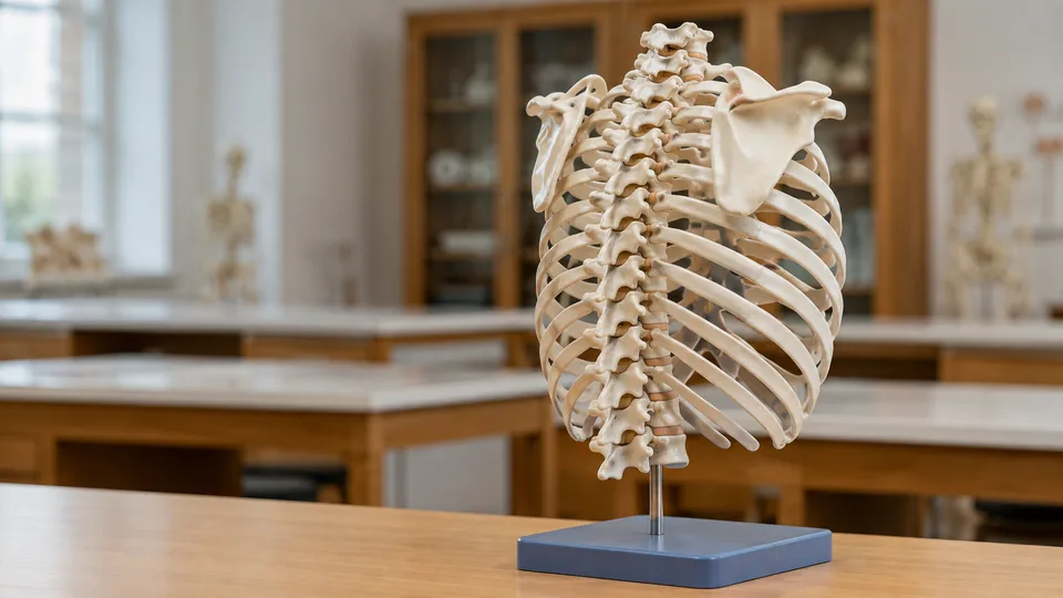
The thoracic spine is easy to underestimate on a printed page. Twelve vertebrae, twelve pairs of ribs, overlapping facets, long spinous processes, and a changing relationship with the cervical and lumbar regions quickly turn a tidy diagram into a visual puzzle. A physical model lets students rotate the anatomy, trace the rib attachments, and see why a thoracic vertebra looks and behaves differently from its neighbors.
Products sold as thoracic spine models vary widely. Some isolate all 12 thoracic vertebrae for focused study, while others place the region inside a flexible vertebral column, rib cage, or cervicothoracic assembly. Buyers often choose a dramatic life-size model before checking whether they need detachable vertebrae, color coding, rib articulation, portability, or a stable tabletop base. Those practical differences matter once the model reaches a crowded study desk or a busy classroom.
For a versatile full-spine reference, the hBARSCI Flexible Human Spine Model is the strongest overall choice here. Its color-coded regions, realistic bony landmarks, pelvis, flexible construction, and hanging stand suit broad anatomy instruction. Learners focused strictly on T1 through T12 should look closely at the dedicated 12-section Thoracic Vertebrae Skeletal Model instead.
Short on reading time? The quick picks and comparison table below show how the leading options differ.
Quick Answer
Best overall: The hBARSCI Flexible Human Spine Model combines life-size regional anatomy, color coding, realistic landmarks, flexibility, and a complete pelvis. Best focused thoracic model: The Thoracic Vertebrae Skeletal Model isolates all 12 thoracic vertebrae and uses detachable segments for close study. Best for rib relationships: The Life-Size Human Rib Cage Anatomy Model reveals how the thoracic spine, ribs, sternum, lumbar region, and pelvis fit together.
Best Thoracic Spine Models Comparison Table
| Product | Best For | Coverage | Key Feature |
|---|---|---|---|
| hBARSCI Flexible Human Spine Model | Overall anatomy instruction | Full spine and pelvis | Flexible, color-coded regions |
| Thoracic Vertebrae Skeletal Model | Focused T1 through T12 study | 12 thoracic vertebrae | Detachable segments |
| Life-Size Rib Cage Model | Thoracic cage relationships | Rib cage, spine, and pelvis | Life-size ribs and flexible sternum |
| breesky Color-Coded Flexible Spine | Beginners and patient education | Full spine and male pelvis | Distinct regional colors |
| breesky Life-Size Spine Model | Flexible movement demonstrations | Full spine and male pelvis | Positionable core and metal stand |
| NLShan Colored Spine Model | Visual teaching and study | Full spine with pelvis | Colored structures and removable femur |
| EVOTECH SCIENTIFIC Neck Vertebrae Model | Cervicothoracic anatomy | Cervical and upper thoracic region | Nerves, arteries, muscles, and brainstem |
| Axis Scientific Vertebrae Set | Compact regional comparison | Representative spine regions | Life-size parts on a tabletop base |
| 3B Scientific 5 Vertebrae | Individual vertebra comparison | Five representative vertebrae | Loosely threaded, portable set |
The detailed reviews below explain where each model shines and which compromises matter in a real study or teaching space.
1. hBARSCI Flexible Human Spine Model With Pelvis: Best Overall

Regional orientation becomes much easier when cervical, thoracic, and lumbar vertebrae are visually separated. The hBARSCI model uses color coding across those three sections while retaining the occipital plate, sacrum, coccyx, and complete pelvis. At 31.5 inches tall, it provides a substantial life-size reference without reducing the thoracic region to an isolated stack of unfamiliar bones.
Its flexible construction supports demonstrations of vertebral movement, curvature, disc relationships, nerves, and arteries. Picture a chiropractic instructor bending the column slightly as a student follows the thoracic curve with one finger. That tactile, three-dimensional lesson lands differently from tracing a line in an atlas. The model is cast from a human skeleton, according to the listing, giving its textures and bony landmarks a realistic appearance that rewards close observation.
Pros
- Color-coded cervical, thoracic, and lumbar regions are easy to distinguish.
- Life-size construction supports clear classroom and clinical demonstrations.
- Flexible column helps illustrate spinal curves and movement relationships.
- Includes an occipital plate, sacrum, coccyx, complete pelvis, nerves, and arteries.
- Washable plastisol polymer and an included hanging stand suit repeated use.
Cons
- The full-spine format is less focused than a detachable T1 through T12 model.
- A 31.5-inch hanging model requires more storage and display room than a desktop set.
This is the most balanced pick for instructors, chiropractic students, physical therapy learners, and clinics that need thoracic anatomy in whole-spine context. Select the dedicated 12-vertebra model below if removing and inspecting individual thoracic levels is the main goal. For broad demonstrations, the hBARSCI model offers the richer all-purpose package.
To buy this product, click here.
2. Thoracic Vertebrae Skeletal Model, 12 Sections: Best Focused Thoracic Model

This model goes straight to the anatomical target. Its 1:1-scale design represents all 12 thoracic vertebrae, avoiding the visual distraction of a skull, pelvis, or long lumbar section. Detachable segments support hands-on inspection, so a learner can study the contours and processes from angles that remain hidden when vertebrae are fixed tightly together.
The listing describes clearly labeled bone and colored detailing, useful when a study partner calls out a structure during a practical-exam rehearsal. Its lightweight PVC construction also makes it easier to carry between a dorm room, laboratory, and skills class. An included instruction manual adds a starting point for beginners, although formal course materials should remain the primary reference for anatomical terminology and identification.
Pros
- Represents all 12 thoracic vertebrae at 1:1 scale.
- Detachable segments encourage close inspection from multiple angles.
- Focused format avoids unrelated full-spine anatomy during thoracic study.
- Colored detailing and labels assist visual identification.
- Lightweight PVC construction is practical for transport and repeated handling.
Cons
- It lacks the complete rib cage and pelvis context provided by larger models.
- Its specialized coverage is less useful for comparing thoracic vertebrae with the entire cervical and lumbar regions.
Medical, massage, osteopathy, and chiropractic students who specifically need T1 through T12 will get more focused value here than from a general spine model. It also suits one-to-one teaching where parts can be passed across a desk. Instructors presenting to a large room may prefer a full-size hanging spine or rib cage assembly with a broader silhouette.
To buy this product, click here.
3. Life-Size Human Rib Cage Anatomy Model With Spine and Pelvis: Best for Thoracic Cage Context

The thoracic spine makes far more spatial sense once the ribs enter the picture. This life-size assembly shows the rib cage alongside the thoracic and lumbar spine and pelvic girdle. From behind, learners can study the vertebral column. Turn it around, and the sweeping ribs and sternum reveal the shape of the thoracic cage in a way an isolated column cannot match.
A soft, compressible sternum permits demonstrations of chest movement and chest-compression mechanics. That feature gives nursing and emergency-education instructors a useful bridge between skeletal anatomy and basic movement concepts, though this remains an educational anatomy model rather than a substitute for a purpose-built CPR manikin. The molded PVC components have smooth contours, and a metal stand supports upright display during teaching.
Pros
- Life-size rib cage clearly shows the thoracic spine within the chest framework.
- Includes thoracic and lumbar spine regions plus the pelvic girdle.
- Flexible sternum supports demonstrations of chest movement.
- Metal stand allows upright classroom or laboratory display.
- Well suited to nursing, physiotherapy, anatomy, and emergency-education settings.
Cons
- The broad skeletal assembly takes up considerably more room than an isolated vertebra set.
- Individual thoracic vertebrae are less accessible than those on a detachable sectional model.
Choose this model when rib articulation, chest shape, and the relationship between the thorax and lower trunk matter most. It can turn an abstract lecture about costovertebral relationships into a clear visual moment across the room. Students concentrating on individual vertebral landmarks will gain more from the focused 12-section model, which allows closer manipulation.
To buy this product, click here.
4. breesky Color-Coded Flexible Spine Model: Best for Beginners

Color can provide the first foothold when a wall of white vertebrae starts to blur together. This 34.6-inch breesky model distinguishes spinal regions with color and depicts vessels and nerves in contrasting tones. The life-size assembly includes 24 vertebrae, the occipital bone, sacrum, coccyx, male pelvis, pubic bone, and femoral heads, creating a broad reference for foundational study.
The flexible column lets an instructor position the spine while discussing regional curves and vertebral relationships. Several pelvic components and the femoral heads can be disassembled, according to the listing, adding useful interaction below the lumbar column. In a patient-education conversation, the bright regional boundaries also make phrases such as “mid-back” more concrete without burying the viewer in tiny labels.
Pros
- Color-coded spinal regions support quick cervical, thoracic, and lumbar orientation.
- Life-size 34.6-inch format provides a clear visual reference.
- Flexible construction helps demonstrate curves and general spinal movement.
- Includes spinal nerves, arteries, pelvis, sacrum, coccyx, and occipital bone.
- Washable PVC and an included stand suit classrooms and offices.
Cons
- Strong color coding looks less bone-like than a neutral model cast for realism.
- The complete column offers less direct access to individual thoracic vertebrae than a detachable set.
This is a sensible choice for first-year students, patient educators, and instructors who value immediate regional recognition. Advanced learners seeking subtle surface detail may favor the hBARSCI model, while thoracic-only study points toward the 12-section pick. For introductory demonstrations, however, the visual separation is wonderfully clear from the first glance.
To buy this product, click here.
5. breesky Life-Size Flexible Spine Model: Best Value Full-Spine Option

For buyers who want a large, positionable column without making vivid regional coloring the centerpiece, this breesky model covers the essentials. It measures 34.6 inches and depicts vertebral bodies, spinous and transverse processes, vertebral notches, the spinal canal, facet joints, nerve branches, and the vertebral artery. The complete pelvis, sacrum, occipital bone, and removable femoral heads broaden its teaching range.
A flexible internal wire allows bending, twisting, and rotation, then holds the selected position for demonstration. That is handy during a tutoring session when one person needs both hands free to point at thoracic landmarks. A lumbar disc feature between L4 and L5 adds another discussion point, although it sits outside the article’s main thoracic focus. The metal stand provides firmer support than a loose model draped across a desk.
Pros
- Life-size column displays the thoracic region within complete spinal anatomy.
- Flexible wire permits bending, rotation, and position holding.
- Shows vertebral landmarks, nerves, an artery, facet joints, and spinal canal.
- Includes a complete pelvis, sacrum, occipital bone, and removable femoral heads.
- Washable PVC construction comes with a supportive metal stand.
Cons
- The featured disc herniation is lumbar rather than thoracic.
- Regional boundaries are less immediately obvious than on a strongly color-coded spine.
This option fits independent learners, small teaching rooms, and offices that need an adaptable full-spine visual aid. Buyers who repeatedly explain cervical, thoracic, and lumbar divisions may prefer the color-coded breesky version. Those who prioritize movement demonstrations and a metal support system should find this simpler design appealing.
To buy this product, click here.
6. NLShan Full-Size Colored Spine Model: Best for Visual Demonstrations

The NLShan model blends a 34.6-inch full-size format with colored structures intended to make anatomical distinctions easier to explain. Nerves, arteries, the lumbar column, and a removable femoral component are included, giving teachers several visible reference points as they move from the thoracic region toward adjacent structures.
Its flexible form is useful for general demonstrations, while the listing describes the vertebral shapes as cast from a human specimen to preserve bony detail. Imagine an art student sketching the sweep of the thoracic curve, then walking around the stand to check the profile from behind. That combination of recognizable landmarks and colorful cues can serve visual learners well, especially during small-group instruction or patient-facing explanations.
Pros
- Full-size 34.6-inch construction offers a substantial visual reference.
- Colored structures help beginners distinguish anatomical components.
- Depicts nerves, arteries, and vertebral landmarks.
- Flexible design supports general spinal positioning demonstrations.
- Removable femoral component adds interaction around the pelvic region.
Cons
- Its broad full-spine coverage leaves the thoracic vertebrae less isolated for detailed inspection.
- Buyers needing a neutral bone appearance may prefer a model with subtler coloring.
Visual learners, anatomy artists, and educators explaining broad spinal organization are the natural audience. It overlaps with other flexible full-spine models, so the choice comes down to presentation style. Pick this one for its mix of color and realistic vertebral contours. Choose the dedicated thoracic model where detachable T1 through T12 study carries more weight.
To buy this product, click here.
7. EVOTECH SCIENTIFIC Life-Size Human Neck Vertebrae Model: Best for the Cervicothoracic Junction

The transition from the mobile cervical spine to the upper thoracic region deserves closer attention than a long hanging column always permits. EVOTECH SCIENTIFIC concentrates on that area with seven cervical vertebrae, upper thoracic vertebrae, spinal nerves, vertebral arteries, surrounding muscles, and the brainstem. Color coding separates arteries, nerves, muscles, and neural tissue at a glance.
Because the assembly sits on a stable white base, it works well during a desk-side explanation where teacher and learner can circle the model without wrestling with a swaying stand. The anatomical scope is specialized. It will not carry a learner through all 12 thoracic levels, yet it offers more soft-tissue and neurovascular context around the upper spine than the skeletal-only picks.
Pros
- Shows the cervical spine together with upper thoracic vertebrae.
- Includes spinal nerves, vertebral arteries, muscles, and brainstem.
- Color coding makes different tissue categories easy to identify.
- Life-size detailing supports close tabletop instruction.
- Stable base is convenient for clinics, classrooms, and study desks.
Cons
- Coverage is limited to the upper thoracic region rather than all 12 thoracic vertebrae.
- The added muscles and neural structures may be busier than necessary for basic bone identification.
Select this model for cervicothoracic transition lessons, upper-spine nerve pathways, or posture-focused educational demonstrations. A student preparing to identify every thoracic vertebra will need broader coverage. For close examination of the neck and upper thorax as one connected region, this compact assembly has a distinct purpose that the full columns do not fill as neatly.
To buy this product, click here.
8. Axis Scientific Vertebrae Set: Best Compact Desktop Reference
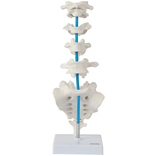
Desk space disappears fast under anatomy atlases, note cards, and a laptop. The Axis Scientific set answers that problem with representative life-size vertebral parts arranged on a 4.25-by-4.25-inch base that stands 13 inches high. It includes the atlas, axis, cervical, thoracic, and lumbar vertebrae, followed by the sacrum and coccyx.
The real advantage is comparison. A student can move visually from the atlas to the thoracic example and down toward the larger lumbar vertebra, observing how regional forms change without scanning a three-foot column. The parts are secured to the base, which keeps the display orderly in an office or study corner. That fixed arrangement trades hands-on disassembly for grab-and-glance convenience.
Pros
- Compact 13-inch display fits comfortably on desks and shelves.
- Includes representative cervical, thoracic, and lumbar anatomy.
- Life-size parts make regional vertebral differences easier to compare.
- Stable base keeps components arranged for quick reference.
- Also includes the atlas, axis, sacrum, and coccyx.
Cons
- A single representative thoracic vertebra cannot show variation across T1 through T12.
- Secured components offer less hands-on manipulation than detachable vertebrae.
This set suits students, professors, and clinicians who need a tidy regional reference rather than a complete thoracic column. It is particularly useful beside a monitor during revision, where a quick sideways glance can settle a regional comparison. Buyers teaching thoracic sequencing, rib attachments, or overall curvature should move toward one of the larger models.
To buy this product, click here.
9. 3B Scientific 5 Vertebrae, A75/1: Best for Individual Vertebra Comparison

Sometimes the quickest route to understanding a thoracic vertebra is placing it beside vertebrae from other regions. This 3B Scientific set contains five examples: atlas, axis, cervical, thoracic, and lumbar vertebrae. They are loosely threaded on nylon, creating a small, tactile comparison set rather than a fixed display tower.
At 0.88 pounds, the group slips into a teaching bag far more easily than a full spine and metal stand. A tutor can pass the set across a table, ask a student to identify the thoracic example, then discuss the landmarks that separate it from the cervical and lumbar pieces. The narrow selection is intentional, but it cannot reveal level-by-level changes through the full thoracic column.
Pros
- Includes representative atlas, axis, cervical, thoracic, and lumbar vertebrae.
- Loose nylon threading allows closer handling and comparison.
- Low 0.88-pound weight makes the set highly portable.
- Focused format works well for quizzes and regional identification practice.
- Registration provides access to the manufacturer’s stated warranty extension.
Cons
- Only one thoracic example is included, so T1 through T12 variation is absent.
- The set lacks ribs, a stand-mounted full column, nerves, vessels, and pelvic context.
Buy this for portable comparative study, tutoring, or quick identification drills. It is far easier to pack than a life-size column and encourages direct handling, which can make regional differences click. Anyone seeking the complete thoracic curve or all 12 vertebrae should treat it as a supplementary reference rather than the primary model.
To buy this product, click here.
How to Choose a Thoracic Spine Model
Decide How Much Anatomy You Need
A dedicated T1 through T12 model is the cleanest option for studying thoracic sequencing and individual vertebral forms. A full-spine model answers a different question: how does the thoracic curve sit between the cervical and lumbar regions? Picture preparing for a practical identification exam versus explaining posture to a patient. The first task rewards detachable thoracic segments, while the second often benefits from a recognizable full column.
Rib cage assemblies add another layer by showing the chest wall around the vertebrae. They are especially useful for nursing, respiratory, physiotherapy, and general anatomy teaching. That context comes with bulk, and the ribs can obstruct close views of some posterior or lateral landmarks.
Check Scale and Viewing Distance
Life-size models are easier to relate to human proportions and typically work well in small-group demonstrations. A tall hanging spine creates a strong silhouette at the front of a room, while a compact vertebra set shines at arm’s length. Think about the actual scene: six students standing around a laboratory bench need a different visual aid from one learner revising beside a laptop.
Dimensions also affect storage. Full columns and rib cages demand vertical clearance, a stand, and a safe corner where projecting parts will not be knocked. Compact sets sacrifice anatomical continuity but can live permanently on a narrow shelf.
Choose Between Flexibility and Detachable Parts
Flexible models help illustrate curves, rotation, and broad movement relationships. Their internal support may allow a pose to be held while the instructor points to a region. Flexibility should be used as an educational approximation, since a model cannot reproduce the full behavior of living joints, muscles, and soft tissues.
Detachable vertebrae serve close structural study. Turning one segment in the hand exposes surfaces hidden inside an assembled spine, but loose pieces require organized storage. Fixed tabletop displays sit between these approaches, offering a tidy comparison while limiting manipulation.
Evaluate Color Coding and Included Structures
Bright regional colors are excellent for beginners and patient education because cervical, thoracic, and lumbar boundaries appear instantly. Neutral bone-colored models often look more realistic and can place greater emphasis on surface contours. Neither approach is universally superior. The right choice depends on whether rapid recognition or bone-like presentation matters more.
Nerves, arteries, muscles, discs, pelvis components, and the occipital bone broaden a model’s teaching range. Extra structures can also create visual clutter during a bone-identification lesson. Pay for the anatomy that supports the intended curriculum rather than the longest feature list.
Look Closely at the Stand and Materials
A stable base matters whenever learners point, rotate, or gather around the model. Hanging stands suit long flexible spines, while weighted tabletop bases work better for compact assemblies. Before ordering, check whether the listed stand fits the available desk height and whether components remain accessible while mounted.
PVC and polymer models are generally practical for repeated educational handling and surface cleaning, following the manufacturer’s care directions. Fine processes and projecting ribs still deserve careful storage. “Durable” should never be read as permission to toss a model loose into a packed equipment bin.
Best Thoracic Spine Models FAQ
Should I Buy a Thoracic-Only Model or a Complete Spine?
Choose a thoracic-only model for concentrated study of T1 through T12, particularly when the vertebrae detach or can be viewed individually. Choose a complete spine for regional orientation, spinal curves, and comparison with cervical and lumbar anatomy. Instructors often gain more versatility from a full column, while exam-focused learners may prefer the isolated thoracic set.
Why Is a Rib Cage Model Useful for Thoracic Spine Study?
The ribs articulate with thoracic vertebrae and form the surrounding thoracic cage. A rib cage model makes those spatial relationships visible, including the shape of the chest and the spine’s position behind it. The trade-off is reduced access to some vertebral surfaces.
Are Color-Coded Spine Models Anatomically Useful?
Yes, particularly for regional recognition and introductory teaching. Color can separate cervical, thoracic, and lumbar sections or distinguish nerves and vessels from bone. Learners preparing to recognize natural bone appearance may also want access to a neutral model or anatomical specimens supplied by their program.
Does a Flexible Spine Model Reproduce Real Human Movement?
It provides a simplified visual demonstration of curves and general movement directions. It does not fully reproduce living joint mechanics, muscular control, ligament behavior, or individual variation. Use flexibility to support educational explanation rather than as a precise biomechanical simulation.
What Thoracic Spine Model Is Best for Medical Students?
The answer depends on the course. A detachable 12-section thoracic model is particularly useful for focused vertebral study. A full-spine model with color-coded regions works better for broad orientation, while a compact comparison set helps learners distinguish typical vertebrae from different regions.
Do I Need Numbered or Labeled Vertebrae?
Labels speed up early learning and self-quizzing, but they can become a crutch if an assessment uses unmarked anatomy. A removable label system or a model paired with a separate identification key offers a useful middle path. Verify listing details carefully, since color coding does not automatically mean every level is numbered.
How Should an Anatomy Model Be Cleaned and Stored?
Follow the manufacturer’s instructions for the specific material. Many PVC or polymer models can be wiped gently, but solvents and abrasive cleaners may damage paint or surfaces. Store the model on its stand or in a protected cabinet, keeping pressure away from ribs, spinous processes, nerves, and other projecting parts.
Final Verdict
Among these nine models, the hBARSCI Flexible Human Spine Model With Pelvis offers the strongest overall balance. Its life-size proportions, color-coded regions, realistic bony landmarks, flexible construction, pelvis, nerves, arteries, and included stand make it useful across classroom instruction, independent study, and patient education. It gives the thoracic region enough visual emphasis while preserving the whole-column context that isolated vertebrae cannot provide.
More specialized buyers have compelling alternatives. The 12-section Thoracic Vertebrae Skeletal Model is the better pick for focused T1 through T12 study and detachable handling. The Life-Size Human Rib Cage Anatomy Model earns its place where ribs, sternum, chest shape, and thoracic spinal relationships drive the lesson. Match the model to the viewing distance, storage space, curriculum, and level of manipulation required. A modest desktop set that answers the right anatomical question is more useful than an elaborate display that never fits the lesson.
