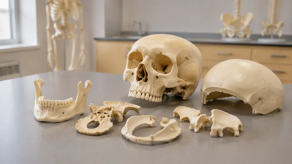
Spread the bones of a human skull across a desk and the anatomy suddenly makes sense in a way a flat diagram rarely can. The sphenoid stops looking like an abstract butterfly, the paired temporal bones reveal their awkward contours, and the facial skeleton becomes a three-dimensional puzzle. Disarticulated skull models are sought by medical and dental students, anatomy instructors, artists, and anyone who needs to see how cranial and facial bones meet beneath the sutures.
The models here differ sharply in scale, component count, color coding, connector design, and anatomical detail. A palm-sized set slips into a backpack but makes tiny foramina harder to inspect. A life-size 22-part skull offers a richer view, though it demands more storage space and patience during assembly. One frequent buying mistake is assuming every “removable” skull fully separates into individual bones. Many affordable models split into only three sections, so the number of removable parts deserves close attention.
For the broadest mix of detail, intuitive assembly, and teaching value, the Axis Scientific 22-Part Didactic Human Skull is the strongest overall choice. Budget-conscious students, traveling tutors, and classroom instructors may prefer very different designs, however, which is why the list spans compact puzzles, life-size models, and one complete osteology set.
Quick Answer
The Axis Scientific 22-Part Didactic Human Skull is the leading option for serious study because its 22 color-coded bones connect magnetically and were cast from a real human skull. The Merinden 22-Part Life-Size Skull offers a more accessible route to full disarticulation, while the 15-Part Palm-Sized Exploded Skull is the easiest model to carry between home, class, and tutoring sessions. For broader osteology study beyond the cranium, the EVOTECH Full-Size Disarticulated Skeleton includes a separate 15-part exploded skull alongside the complete skeletal set.
Short on study time? The table provides a fast look at the differences before the detailed reviews begin.
Best Disarticulated Skull Models Comparison Table
| Product | Best For | Scale or Size | Removable Parts and Key Feature |
|---|---|---|---|
| Axis Scientific Didactic Skull | Overall anatomical study | Life-size | 22 color-coded bones with magnetic connections |
| Merinden Disarticulated Skull | Students seeking full disarticulation | Life-size | 22 color-coded parts and illustrated manual |
| Colored Beauchene Skull | Classroom demonstrations | 7.67 × 5.9 × 5.11 inches | 22 parts in nine colors with stable connectors |
| Axis Scientific 22-Part Skull | Natural-looking anatomical reference | Life-size | 22 detailed components and full-color manual |
| 15-Part Palm-Sized Skull | Portable study | 4.49 × 3.44 × 4.49 inches | 15 colored parts with removable skullcap |
| Half-Size Didactic Skull | Learning through assembly | Half life-size | 15 colored parts and illustrated question guide |
| JENACAD 55-Landmark Skull | Practical-exam review | Life-size | Three parts, 55 numbered landmarks, red sutures |
| Life-Size Three-Part Skull | Budget anatomical reference | Life-size | Removable calvarium and mandible |
| EVOTECH Skeleton and Skull Set | Whole-body osteology | 67-inch skeleton | Complete skeletal set plus 15-part exploded skull |
The reviews below explore how each design behaves as a study tool and where its compromises become noticeable.
1. Axis Scientific 22-Part Didactic Human Skull: Best Overall

Pulling apart a skull bone by bone can turn a murky lecture on cranial anatomy into a satisfying click-by-click lesson. This Axis Scientific model separates into 22 color-coded bones, each joined to its neighbors by magnets. That arrangement reduces the struggle commonly associated with pegs and tight plastic connectors. It is especially useful during repeated drills, where a student might remove the sphenoid, replace it, and then trace its relationships with the temporal and zygomatic bones.
The model was cast from a real human skull, so the listing emphasizes authentic suture lines, foramina, contours, and surface detail rather than a generic approximation. Its accompanying full-color manual identifies more than 50 structures using photographs of the model. That combination suits medical, dental, osteopathic, forensic science, and advanced anatomy settings where subtle landmarks matter as much as naming each bone.
Pros
- Separates into 22 individual cranial and facial bones
- Magnetic joints make repeated assembly relatively intuitive
- Distinct bone colors support rapid visual identification
- Cast from a real human skull for realistic surface anatomy
- Includes a detailed photographic manual covering more than 50 structures
Cons
- Premium construction places it above basic student models in cost
- Numerous small components require organized storage and careful handling
This is the model to choose when skull anatomy will be studied repeatedly rather than displayed occasionally. It brings color, tactile assembly, and strong surface detail together without asking students to wrestle with stubborn connectors. A casual art student may find its 22-piece complexity excessive, but learners preparing for demanding anatomy coursework can extract considerable value from that complexity.
To buy this product, click here.
2. Merinden 22-Part Life-Size Disarticulated Skull: Best Value

Students who want full disarticulation without stepping into the highest product tier should look closely at the Merinden. Its adult life-size form separates into 22 labeled components, allowing the frontal, parietal, temporal, sphenoid, zygomatic, and other bones to be handled independently. Distinct colors make a crowded study table much less intimidating, particularly during the first weeks of cranial osteology.
An illustrated full-color manual maps the pieces and supplies anatomical names, which gives independent learners a reference when an instructor is unavailable. The sturdy, non-toxic plastic construction and secure connectors are intended for recurring assembly, disassembly, and cleaning. Picture a small study group passing the bones around before reconstructing the face. This model supports that sort of tactile collaboration while retaining realistic adult proportions.
Pros
- Provides 22 removable parts at life-size scale
- Color coding clarifies boundaries between neighboring bones
- Includes an illustrated guide for identification and reassembly
- Secure connectors support recurring classroom use
- Suitable for medical, dental, teaching, and display settings
Cons
- Assembly requires more time than a three-part skull
- Colored surfaces look less natural than a bone-tone reference model
The Merinden strikes a practical middle ground for students who need individual-bone study but cannot justify the premium Axis model. Its guidebook and bright visual separation should help beginners work through an initially confusing pile of cranial pieces. Artists seeking a natural bone appearance may prefer a neutral model, while anatomy students focused on relationships between bones are likely to appreciate the bold palette.
To buy this product, click here.
3. 22-Part Colored Beauchene Exploded Skull Model: Best for Classrooms

Across a classroom, pale bones tend to blur into one chalky mass. This Beauchene-style model answers that problem with 22 parts presented in nine colors, giving instructors clear visual boundaries to point toward during a demonstration. The skull measures 7.67 by 5.9 by 5.11 inches, close enough to adult scale for discussing the orbit, cranial cavity, nasal cavity, sutures, canals, and dental arrangement.
The model is described as being cast from a real specimen and includes a complete set of teeth, an articulating mandible, and removable skullcap. Individual bones reconnect along simplified sutures using stable, unobtrusive connectors. Those simplified joins favor practical reassembly over a perfectly seamless exterior, a sensible trade for a teaching model that may be taken apart between lectures and passed around a lab bench.
Pros
- Twenty-two removable components support bone-by-bone instruction
- Nine-color scheme remains easy to interpret during demonstrations
- Life-size proportions provide useful spatial context
- Removable mandible and skullcap expose internal regions
- Durable PVC surface is straightforward to clean
Cons
- Simplified connector lines may look less natural than true sutures
- Nine colors do not give every paired component a completely unique shade
Instructors, tutors, and group-study leaders are the natural audience here. The bright sections make verbal directions such as “follow the temporal bone forward” much easier for learners to follow from a few feet away. Buyers seeking a museum-like bone finish should look elsewhere, but classrooms gain more from immediate visual clarity than subtle cosmetic realism.
To buy this product, click here.
4. Axis Scientific 22-Part Disarticulated Human Skull: Best Natural-Looking Reference

Color coding is brilliant for memorization, yet some learners eventually need to recognize landmarks without a rainbow acting as a hint. This life-size Axis Scientific replica emphasizes natural anatomical textures, bony ridges, and small components such as the nasal bones. All 22 parts can be handled separately, giving students the tactile benefit of full disarticulation with a more realistic visual presentation.
The included full-color manual identifies more than a dozen numbered features. That is less extensive than the guide supplied with the didactic magnetic Axis model, but it still provides a useful starting point for self-directed review. For an art studio, osteology table, or advanced classroom, the neutral appearance also keeps attention on form, contour, and spatial relationships rather than painted categories.
Pros
- Life-size design preserves realistic cranial proportions
- Twenty-two separate parts permit detailed osteology study
- Natural textures and ridges suit visual anatomical reference
- Small facial bones receive dedicated attention
- Full-color manual supports guided identification
Cons
- Neutral coloration offers fewer visual cues for beginners
- Included manual covers fewer labeled features than the premium didactic Axis guide
Choose this Axis model when realistic form carries more weight than fast color-based memorization. It can serve medical learners who have progressed beyond introductory identification and artists studying the planes beneath the face. A first-semester student who feels lost among similar pale pieces may have an easier start with the colored Merinden or Beauchene models.
To buy this product, click here.
5. 15-Part Palm-Sized Human Skull Model: Best Compact Option

A full-size skull can dominate a crowded desk, while this 15-piece version occupies roughly the space of a large mug. Its listed dimensions are 4.49 inches high, 3.44 inches wide, and 4.49 inches long. The colored components reveal major bone groups, and the removable skullcap opens the endocranial area for a closer view of the internal cavity.
Despite its reduced scale, the model depicts eye sockets, sutures, canals, the nasal cavity, an articulating mandible, and a complete set of teeth. Durable PVC keeps it light and washable, qualities that matter when the set travels in a backpack beside notebooks and lab gear. The smaller pieces can feel fiddly in large hands, but the ability to rehearse assembly at a library table or during tutoring is a real advantage.
Pros
- Compact dimensions make the model easy to carry and store
- Fifteen colored parts simplify major bone identification
- Removable skullcap exposes the internal cranial space
- Includes an articulating mandible and full set of teeth
- Washable PVC suits recurring educational use
Cons
- Reduced scale makes small foramina and fine landmarks harder to inspect
- Fifteen-part layout provides less separation than a 22-bone model
This pocketable skull makes sense for commuters, traveling tutors, and students with limited shelf space. It is strongest as a portable supplement for learning major bone relationships rather than a definitive reference for tiny landmarks. Anyone preparing for a detailed cranial-base practical should lean toward life-size anatomy, but casual review becomes far easier when the model can go almost anywhere.
To buy this product, click here.
6. 15-Part Half-Size Didactic Skull Model: Best for Beginners

Beginners often learn faster when assembly feels like a puzzle rather than a high-stakes lab practical. This half-life-size model divides the skull into 15 colored components and comes with an illustrated guide containing general information and study questions. That guided format creates a ready-made routine: identify a piece, locate its neighbors, rebuild the skull, and use the questions to check recall.
The compact skull includes visible sockets, canals, teeth, sutures, the internal nasal cavity, and the endocranial cavity. Its mandible articulates, adding a basic view of how the lower jaw relates to the cranium. It will not deliver the spacious inspection surface of a life-size model, yet its light, manageable format is less daunting for a learner opening an anatomy kit for the first time.
Pros
- Fifteen-piece format is approachable for new anatomy students
- Colored bone groups make initial identification easier
- Illustrated guide includes questions for active recall
- Half-size construction is portable and space efficient
- Shows key cavities, sutures, sockets, and dental anatomy
Cons
- Half-size anatomy limits close inspection of delicate structures
- Grouped 15-part construction is less comprehensive than full 22-part separation
This model is well matched to introductory anatomy courses, homeschool lessons, and learners who value a structured guide. Its educational strength lies in repetition and approachable scale, not in reproducing every bone as a separate life-size object. Advanced dental or medical students may outgrow it, while beginners can enjoy a gentler path into a notoriously complex part of the skeleton.
To buy this product, click here.
7. JENACAD Life-Size Skull With 55 Landmarks: Best for Practical-Exam Review

Full disarticulation is excellent for bone relationships, but many lab practicals ask students to name a pin-marked feature on an assembled skull. JENACAD takes direct aim at that task with 55 numbered landmarks, black identifiers, and red-painted sutures on a bone-white surface. A printed study card, downloadable PDF guide, and guided videos create several ways to check an answer after attempting recall.
This life-size model separates into three sections: a magnetic skullcap, cranial base, and removable mandible. The spring-supported jaw moves to demonstrate tooth position, occlusion, and mandibular relationships, while the removable calvarium exposes internal surfaces. At about 7.5 by 5.5 by 6.5 inches and two pounds, it offers realistic proportions without the loose-piece management required by a 22-part model.
Pros
- Fifty-five numbered landmarks support active self-testing
- Red sutures stand out clearly against the bone-white surface
- Includes printed and digital study resources
- Life-size proportions suit practical-exam preparation
- Magnetic skullcap and movable jaw improve access to key regions
Cons
- Three-part construction does not separate individual cranial bones
- Printed numbers and red sutures reduce natural visual realism
Students revising foramina, sutures, and named landmarks may get more daily utility from this model than from a loose 22-piece puzzle. It is quick to open, quiz, and return to a shelf after a late-night review session. Buyers whose coursework centers on assembling individual cranial bones should select a fully disarticulated option, while practical-focused learners can benefit from its built-in prompts.
To buy this product, click here.
8. Life-Size Three-Part Anatomical Skull Model: Best Budget Life-Size Option

Sometimes the goal is simple: inspect a life-size cranial base, lift off the calvarium, and move the mandible without managing a tray of tiny parts. This PVC model separates into those three core sections. Magnets and small pegs secure the removable calvarium, allowing it to lift away for study and settle back into position without complicated reconstruction.
The listing describes realistic bone texture, clear sutures, and visible foramina and fossae. A colorful skull chart is included for reference, which helps connect the unpainted model with named regions during independent study. Its neutral finish also suits drawing practice, patient education, or a classroom shelf where natural-looking proportions matter more than individual removable bones.
Pros
- Life-size format provides realistic spatial proportions
- Three-part construction is quick to open and reassemble
- Magnetic calvarium combines access with secure placement
- Neutral bone finish works well for art and general reference
- Includes a colorful anatomical chart
Cons
- Cranial and facial bones cannot be removed individually
- Lacks built-in numbers or color coding for rapid identification
This is a sensible entry point for buyers who need an assembled skull with access to its interior rather than true bone-by-bone disarticulation. It is easier to live with on a cramped desk and faster to use during a short review break. Students specifically assigned to identify isolated sphenoid, ethmoid, or palatine bones will need one of the 22-part selections instead.
To buy this product, click here.
9. EVOTECH 67-Inch Disarticulated Skeleton With Exploded Skull: Best for Whole-Body Osteology

Cranial anatomy rarely stays isolated for long. Before long, a course moves down to vertebrae, ribs, the pelvis, and the wonderfully chaotic bones of the hands and feet. This EVOTECH package supports that broader path with a 67-inch disarticulated PVC skeleton plus a separate half-size, 15-part colored exploded skull. The skull depicts major sutures, cavities, sockets, textures, and an articulating mandible.
The skeletal collection includes a spine with cartilage connections, pelvic components, intervertebral discs, hyoid, coccyx, and articulated versions of the left hand and foot. A laminated identification poster accompanies the set. This is a sprawling study kit rather than a tidy desktop purchase, but it lets an osteology student compare cranial pieces with bones from the rest of the body in the same session.
Pros
- Includes a separate 15-part colored exploded skull
- Expands study across a complete full-size skeletal collection
- Articulated hand and foot preserve small-bone relationships
- Includes structures such as the hyoid, coccyx, and intervertebral discs
- Laminated poster provides a convenient identification reference
Cons
- Requires substantially more storage and sorting space than a skull-only model
- The included skull is half-size rather than a full-size 22-part specimen
This set fits osteology labs, physical anthropology study, artists building a whole-body reference library, and students whose coursework extends far beyond the skull. It would be excessive for someone focused solely on cranial anatomy, and loose skeletal pieces demand careful organization. For broad anatomy study, however, the combination offers a scope none of the skull-only picks can match.
To buy this product, click here.
How to Choose a Disarticulated Skull Model
Start With the Number of Removable Parts
The phrase “disarticulated skull” covers very different constructions. A 22-part model generally separates the major cranial and facial bones, making it possible to hold the sphenoid or temporal bone independently and inspect surfaces hidden in an assembled skull. A 15-part model usually groups some paired or neighboring structures, which makes assembly simpler but limits bone-by-bone study. Three-part skulls remove only the calvarium and mandible from the cranial base.
Match that component count to the assignment. A dental student tracing the pterygopalatine region may benefit from full separation, while a nursing student reviewing major sutures and foramina may work faster with a three-part numbered model. More pieces create more learning possibilities, yet they also create a bigger sorting and storage job.
Choose Between Life-Size and Compact Anatomy
Life-size models provide the clearest view of small openings, fossae, ridges, and internal surfaces. They also give artists and healthcare students a realistic sense of proportion. During a group lesson, a larger skull is easier to point at and pass around without everyone leaning across the table.
Compact models trade inspection space for portability. A half-size skull can travel in a backpack and fit beside a laptop in the library. Tiny components are easier to misplace, however, and subtle landmarks may be difficult to distinguish. Use compact anatomy for major relationships and repetition, then consult a life-size reference for fine detail.
Color Coding Versus Natural Bone Color
Bright colors reduce the mental load of distinguishing tightly packed bones. The boundary between the sphenoid, temporal, and zygomatic bones becomes immediate rather than something learners must hunt for along pale sutures. This helps during introductory lessons and room-wide demonstrations, where a vivid bone is easier to identify from a distance.
Natural bone-tone models better resemble specimens and practical-exam materials. They also serve artists who want to study form without painted visual cues. A useful progression is to begin with color coding, then shift toward a neutral model or cover the color key once identification becomes comfortable.
Examine the Connector System
Magnets are quick and satisfying, producing a clean snap as neighboring components meet. They work particularly well for repeated reconstruction drills. Pegs and fitted connectors can also hold securely, though tight joints may require more deliberate handling. Simplified suture connections favor convenience but may interrupt the natural appearance of the exterior.
No connector system makes delicate anatomy indestructible. Small facial bones, thin projections, and tiny pieces deserve a divided storage container rather than a loose tote bag. Before buying for a shared classroom, consider who will manage the components and how often the skull will be rebuilt.
Look Closely at Study Materials
A detailed guide can turn a pile of colorful plastic into an organized lesson. Photographic manuals are especially useful because the pictured textures and colors match the model in front of the learner. Numbered landmarks support self-quizzing, while question sheets encourage active recall instead of passive viewing.
Check whether the learning materials match the required curriculum. A brief chart may cover major bones yet omit the foramina, processes, and fossae expected in an advanced course. Product guides should supplement lectures, atlases, and formal instruction rather than replace them.
Plan for Storage and Classroom Use
Twenty-two loose pieces have a habit of migrating across desks. A labeled compartment box, soft mat, and written inventory can prevent a tiny nasal or lacrimal component from vanishing after group study. Washable PVC is convenient in shared settings, but the manufacturer’s care directions should guide cleaning and handling.
Instructors also need visibility. A colored life-size model works well at the front of a small classroom, while a compact set is better passed between two or three learners. Whole-skeleton packages need bins, shelves, and considerable floor or table space, so measure the storage area before a very large carton arrives.
Best Disarticulated Skull Models FAQ
How Many Pieces Should a Disarticulated Skull Have?
A 22-part model is the strongest choice for studying major cranial and facial bones individually. Fifteen-part skulls group some anatomy and work well for portable or introductory study. Three-part models offer access to the cranial interior but do not separate individual bones.
Are Color-Coded Skull Models Anatomically Useful?
Yes. Color coding makes adjacent bones easier to distinguish and supports fast visual recall, especially for beginners. The colors are instructional rather than natural, so advanced students may also want exposure to neutral bone-tone models before specimen-based assessments.
Is a Half-Size Skull Large Enough for Medical Study?
A half-size skull is useful for learning major bones, cavities, and spatial relationships. It is less suitable for close inspection of small foramina, delicate processes, and subtle surface landmarks. Life-size anatomy is generally more practical for detailed medical or dental study.
What Is the Difference Between a Three-Part and 22-Part Skull?
A three-part skull typically divides into the calvarium, cranial base, and mandible. A 22-part model separates the major individual bones, allowing learners to examine hidden surfaces and reconstruct their relationships. The 22-part design provides greater depth but takes longer to assemble.
Are Magnetic Skull Models Better Than Peg-Connected Models?
Magnetic models tend to be faster and more intuitive to assemble, which suits frequent study drills. Peg-connected models can also be secure and effective, though alignment may require more care. Anatomical detail, scale, and component design matter as much as the connector type.
Can Disarticulated Skull Models Be Used for Art Reference?
Yes. Life-size models help artists understand cranial proportions, facial planes, orbital depth, and the bony structure beneath soft tissue. Neutral bone-tone skulls usually provide the cleanest visual reference, while colored models are helpful for learning which bone creates each visible contour.
How Should Loose Skull Components Be Stored?
Keep the pieces in a divided container with a simple inventory list. A padded or lined box protects projections from knocking together, and labels make missing components easier to spot. Store the model away from excessive heat and follow the maker’s care instructions for cleaning.
Final Verdict
The Axis Scientific 22-Part Didactic Human Skull earns the top position because it combines life-size anatomy, 22 color-coded bones, magnetic assembly, real-skull casting, and a detailed photographic manual. It is a compelling fit for serious medical, dental, osteopathic, and anatomy study where students need to move repeatedly between isolated bones and a complete cranium. The premium tier will be excessive for casual reference, but its thoughtful connector system helps make complex anatomy feel far less clumsy.
For a more accessible fully disarticulated choice, the Merinden 22-Part Life-Size Skull delivers color-coded components and an illustrated manual in a classroom-ready format. Travelers and students in tight living spaces should consider the 15-Part Palm-Sized Skull, while learners preparing for numbered practicals may gain more from the JENACAD 55-Landmark Skull. Before purchasing, check the course objectives, available storage, and required level of detail. The right model is the one that makes the assigned anatomy visible, repeatable, and easy to revisit at the study desk.
