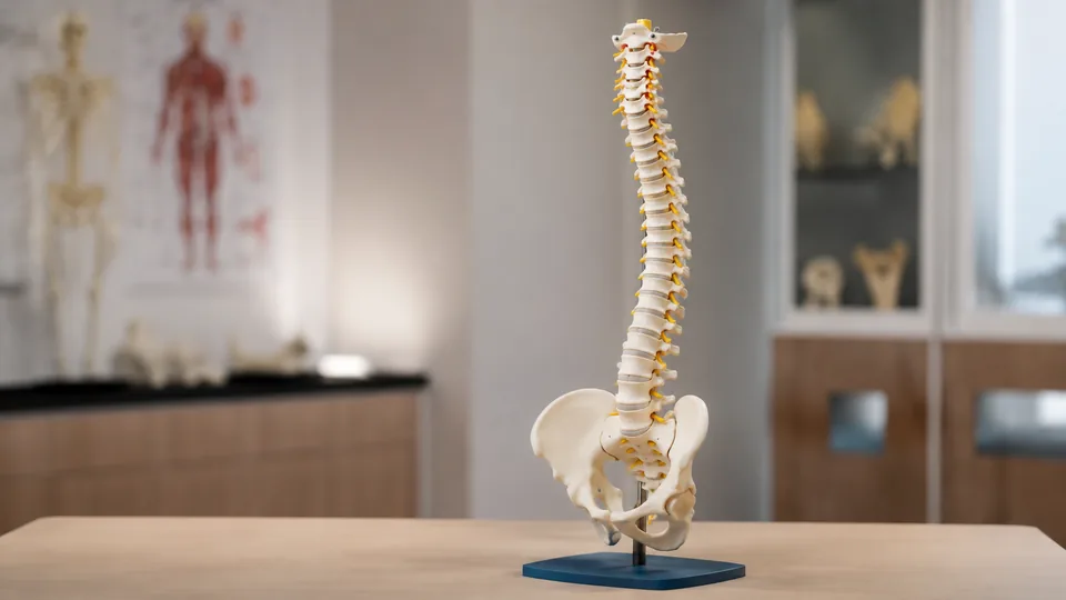
A spine model can turn a flat textbook diagram into something instantly understandable. Rotate the pelvis, follow a nerve branch, bend the vertebral column, and the anatomy suddenly has shape, depth, and context. The best spine models with pelvis are useful for anatomy students, physical therapy programs, chiropractic classrooms, patient education, art reference, and anyone who needs to see how the vertebral column connects with the pelvic girdle.
Models in this category differ more than their white plastic bones suggest. Some emphasize flexible movement, while others add color coding, removable femur heads, muscle markings, nerves, arteries, or a wider view of the rib cage. A common buying mistake is choosing by height alone. Scale matters, but anatomical detail, stand stability, flexibility, and the intended lesson matter far more. The breesky Spine Model offers the strongest overall balance here, pairing life-size anatomy with a flexible core, male pelvis, removable femur heads, and a sturdy display stand.
Different settings call for different priorities. A student studying vertebral landmarks may prefer clear color differentiation, while an instructor working across a large room needs a full-size model that stays upright during demonstrations. Someone teaching female pelvic anatomy needs a fundamentally different design from a buyer focused on the entire vertebral column, which is where this list begins to spread out.
Quick Answer
Best overall: The breesky Spine Model combines life-size proportions, flexible positioning, nerves, arteries, a lumbar disc herniation, and a complete pelvis. The 34.6-Inch Life-Size Spine Model is the value-oriented choice, especially for buyers who appreciate the included anatomy chart. For a broader view connecting the spine, pelvis, and thorax, the Life-Size Human Rib Cage Anatomy Model provides useful whole-region context.
Short on reading time? The table below shows how the nine selections differ at a glance.
Best Spine Models With Pelvis Comparison Table
| Product | Best For | Scale or Size | Key Feature |
|---|---|---|---|
| breesky Spine Model | Most buyers | 34.6 inches, life size | Flexible spine with removable femur heads |
| 34.6-Inch Life-Size Spine Model | Value and guided study | 34.6 inches | Includes an anatomy chart |
| Axis Scientific Spine Model | Classrooms and clinics | 34 inches, life size | Full-color product manual |
| JNMFTD Female Pelvis Model | Female pelvic anatomy | 1:1 life size | Muscles, nerves, hip joints, and movable femur heads |
| NLShan Colored Spine Model | Visual learners | 34.6 inches, full size | Color-differentiated bone structures |
| Rib Cage, Spine, and Pelvis Model | Thoracic context | 1:1 life size | Rib cage and compressible sternum |
| Flexible Male Pelvis Spine Model | Movement demonstrations | 34.6 inches, life size | Flexible column and metal support |
| Colorful Spine Model | Patient explanations | 34.6 inches, life size | Colored anatomy with visible nerves and arteries |
| 3B Scientific Flexible Spine Model | Premium instructional use | Life size | Functional flexibility with pelvis and femur heads |
The detailed reviews below explain where each model fits, from solo study at a cramped desk to demonstrations in a busy anatomy lab.
1. breesky Spine Model: Best Overall

The breesky model earns the top position by covering the details most buyers actually use. Its 34.6-inch vertebral column shows vertebral bodies, spinous and transverse processes, facet joints, the spinal canal, nerve branches, and a vertebral artery. A represented herniated disc between L4 and L5 gives instructors a visible reference for a condition that can feel frustratingly abstract on a printed page. The complete male pelvis, sacrum, occipital bone, and removable femur heads extend the lesson beyond an isolated stack of vertebrae.
A flexible internal wire allows the column to bend, twist, rotate, and hold a selected position. Picture an instructor demonstrating lateral flexion, then stepping aside so students can inspect the altered curves from several angles. That tactile, three-dimensional view is the model’s big draw. The PVC construction is washable, while the metal stand and base keep the long model displayed upright between lessons. It suits classrooms, physical therapy instruction, chiropractic education, patient conversations, and artists studying posture.
Pros
- Life-size spine includes a complete male pelvis and sacrum
- Flexible core supports bending, twisting, and rotational demonstrations
- Shows nerves, a vertebral artery, facet joints, and an L4-L5 disc herniation
- Removable femur heads help illustrate the pelvis-to-hip relationship
- Metal stand provides convenient upright storage and display
Cons
- Its full length requires more display and storage space than a lumbar-only model
- Male pelvic anatomy does not serve lessons focused specifically on female pelvic structure
This is the practical all-rounder for buyers who want one model to handle vertebral identification, movement demonstrations, and basic pelvic orientation. A specialized lab may need finer regional models alongside it, but students, instructors, and patient educators should find its combination of flexible movement and visible structures unusually useful. Buyers with limited shelving should measure first, since a 34.6-inch model plus its support makes a substantial presence in a small room.
To buy this product, click here.
2. 34.6-Inch Life-Size Spine Model: Best Value

This 34.6-inch model is a compelling value choice because it combines full-size anatomy with a study chart. The physical model depicts vertebral bodies, spinous and transverse processes, the spinal canal, small joints, nerve branches, vertebral arteries, and a disc herniation at L4-L5. It also includes the pelvis, sacrum, and occipital bone, giving students a continuous view from the base of the skull through the pelvic region.
The flexible construction is useful during lessons on spinal curvature, flexion, extension, and rotation. A student can read about lumbar movement on the supplied chart, then immediately shape the model and inspect how the curves relate to the pelvis. The base covers approximately 7 by 7 inches, and a metal support helps the tall structure remain upright. That combination balances a manageable footprint with enough height for visible tabletop or classroom demonstrations.
Pros
- Life-size format supports realistic spatial study
- Flexible column demonstrates several directions of movement
- Includes nerves, arteries, small joints, and an L4-L5 disc herniation
- Supplied anatomy chart offers a convenient secondary reference
- Metal support and compact base aid upright display
Cons
- The listing provides less detail about removable components than some competing models
- Full-size construction can be cumbersome for students who commute with their supplies
Buyers assembling a home study corner or equipping a teaching room on a moderate budget should give this model a close look. The chart is especially handy for beginners who still need names beside the shapes they are handling. Experienced instructors seeking extensive muscle markings or a highly specialized pelvic model should look elsewhere, but this option covers the core spine-and-pelvis lesson without unnecessary fuss.
To buy this product, click here.
3. Axis Scientific 34-Inch Life-Size Spine Model: Best for Classrooms

Axis Scientific packages its 34-inch model as a broad educational resource rather than a bare display piece. The life-size vertebral column includes the lumbar region, individual vertebrae, spinal nerves, arteries, and a male pelvis. Its scale works well when a teacher needs to point out a landmark to several people gathered around a lab table, yet it remains manageable enough for individual study after class.
The flexible spine supports demonstrations of natural curves and basic movement. A colorful manual adds another route into the material, which can be useful during a first anatomy course when every process and foramen seems to blur together. The included stand also matters in a classroom. Instead of laying the model across worksheets and laptops, an instructor can keep it upright, visible, and ready for the next group.
Pros
- Life-size 34-inch design is suitable for group demonstrations
- Includes spinal nerves, arteries, lumbar vertebrae, and male pelvis
- Flexible construction supports movement and curvature lessons
- Durable stand keeps the model upright during display
- Full-color manual assists structured study and review
Cons
- Color-coded bone regions are less prominent than on models created specifically for visual differentiation
- Buyers focused on female pelvic anatomy will need a different anatomical configuration
This model makes the most sense in classrooms, chiropractic offices, and shared study spaces where a recognizable full-size reference may move between several users. The manual gives beginners a useful starting point, while the stand makes daily storage tidier. Buyers who need muscles or movable hip joints should favor the JNMFTD model, but for a classic spine, nerves, and pelvis presentation, Axis Scientific is a polished choice.
To buy this product, click here.
4. JNMFTD Female Pelvis and Lumbar Spine Model: Best for Female Pelvic Anatomy

The JNMFTD takes a regional approach, placing far more emphasis on the female pelvis, hip joints, and lumbar connection than a conventional full-column model. Its 1:1 anatomy includes the lumbar spine, pelvis, femurs, nerves, and muscle features. Movable, removable femur heads let learners inspect how the proximal femur meets the acetabulum, which is much easier to grasp when the parts can be repositioned in front of you.
Colored structures create visual separation across an area where bone, nerves, and muscular anatomy can quickly become crowded. A detailed product manual supports identification, while the removable base gives users a choice between stable display and closer handling. Imagine a small-group lesson focused on pelvic landmarks and hip orientation. This model puts that region at center stage rather than leaving it dangling beneath a long vertebral column.
Pros
- Represents female pelvic anatomy at 1:1 scale
- Includes lumbar spine, nerves, muscles, pelvis, and hip structures
- Movable and removable femur heads support hip-joint demonstrations
- Colored features make neighboring structures easier to distinguish
- Includes a manual and removable sturdy base
Cons
- Lumbar-focused design does not show the complete cervical and thoracic spine
- Greater regional detail may be unnecessary for basic vertebral-column lessons
Choose this model for coursework or teaching centered on the female pelvis, lumbar region, and hip relationship. It is also a strong visual reference for artists trying to understand how pelvic structure influences stance. Anyone seeking a continuous occiput-to-pelvis model should skip it, since the regional format deliberately trades total spinal coverage for deeper pelvic context.
To buy this product, click here.
5. NLShan Colored Full-Size Spine Model: Best for Visual Learners

A white-on-white anatomical model can become a maze under fluorescent classroom lights. NLShan addresses that problem by painting different bone structures in contrasting colors, helping beginners separate regions and landmarks at a glance. The 34.6-inch model includes the vertebral column, nerves, arteries, lumbar region, pelvis, and removable femur components, making it useful for broad survey courses and patient-friendly explanations.
The listing describes a flexible construction based on details captured from human spinal bones. That flexibility lets an instructor position the column while discussing posture or basic biomechanics, while the color treatment helps an observer keep track of the structures being referenced. PVC construction also makes the model relatively straightforward to wipe clean after repeated handling in a lab or classroom, where fingerprints and pencil smudges seem to appear from nowhere.
Pros
- Contrasting colors help distinguish anatomical regions
- Full-size 34.6-inch format offers clear classroom visibility
- Includes nerves, arteries, lumbar column, and pelvis
- Flexible spinal construction supports positional demonstrations
- Removable femur components add pelvic and hip context
Cons
- Strong color coding looks less naturalistic than plain bone-colored models
- Buyers who already know the regional anatomy may gain less from the painted distinctions
NLShan is particularly appealing for introductory students, patient education, and visual learners who benefit from immediate separation between structures. Its bright treatment can make a spoken explanation easier to follow from across a table. Medical illustrators seeking natural bone coloration may prefer Axis Scientific or breesky, but for clarity during a fast-moving lesson, the colors can earn their keep.
To buy this product, click here.
6. Life-Size Human Rib Cage Anatomy Model With Spine and Pelvis: Best for Thoracic Context

This model zooms out. Rather than presenting the spine as a lone vertical column, it connects the thoracic and lumbar regions with a life-size rib cage and pelvic girdle. That wider skeletal context is valuable for nursing, physiotherapy, emergency education, and anatomy classes where learners need to understand how the vertebrae sit behind the ribs and above the pelvis. Smooth molded contours and PVC construction support repeated educational handling.
The soft, compressible sternum is its most distinctive feature. It can be used to demonstrate the anatomical mechanics associated with chest compression and respiratory movement without pretending to replace a dedicated training manikin. Picture an instructor tracing the thoracic spine, moving forward along a rib, and then pressing the sternum to connect posterior and anterior anatomy. A metal stand holds the large assembly upright so that the full relationship remains visible.
Pros
- Shows the rib cage, thoracic and lumbar spine, and pelvis together
- Life-size scale provides realistic skeletal proportions
- Compressible sternum supports basic chest-mechanics demonstrations
- Durable PVC construction suits repeated classroom handling
- Metal stand provides stable upright presentation
Cons
- The broad torso format takes up more room than a spine-only model
- It offers less emphasis on isolated nerves and vertebral arteries than several dedicated spine models
This is the better choice for buyers teaching regional relationships across the torso. Nursing and emergency education programs may particularly appreciate seeing the rib cage, sternum, spine, and pelvis as one connected structure. Students preparing for detailed identification of nerve branches or vascular features should choose a dedicated spine model instead, since the strength here is anatomical context rather than tiny isolated detail.
To buy this product, click here.
7. 34.6-Inch Flexible Spine Model With Male Pelvis: Best for Movement Demonstrations

For instructors who repeatedly demonstrate bending, twisting, flexion, and extension, this 34.6-inch model puts flexibility front and center. It includes vertebral bodies, processes, the spinal canal, joints, nerve branches, vertebral arteries, an L4-L5 disc herniation, sacrum, occipital bone, and a complete male pelvis. The result is a familiar life-size teaching format with enough added detail to support discussions beyond simple bone identification.
The flexible column can be shaped to illustrate different curves and movement directions, then returned to its display position. A sturdy metal support helps protect the long assembly from accidental tipping, which matters when several students crowd around and someone inevitably nudges the table. Its washable, lightweight PVC construction also fits environments where the model will be passed from hand to hand rather than sitting untouched behind glass.
Pros
- Flexible spine supports bending, twisting, and rotation lessons
- Life-size format includes a complete male pelvis and occipital bone
- Displays nerves, vertebral arteries, joints, and an L4-L5 disc herniation
- Removable femur heads expand pelvic demonstrations
- Metal support helps stabilize the model during display
Cons
- Frequent repositioning still calls for careful handling of projecting structures
- Its feature set overlaps with other full-size flexible models, offering less specialization
This option suits physical therapy education, chiropractic instruction, and anatomy lessons where movement appears repeatedly in the curriculum. It is less compelling for buyers who need female pelvic structures, muscle markings, or the surrounding rib cage. Among conventional flexible models, however, its complete set of nerves, arteries, pelvis, and represented disc pathology covers the everyday teaching essentials cleanly.
To buy this product, click here.
8. 34.6-Inch Colorful Anatomy Spine Model: Best for Patient Explanations

This colorful model pairs a flexible, full-size vertebral column with anatomy that is easier for a newcomer to follow. It depicts the vertebral bodies and processes, spinal canal, small joints, nerve branches, vertebral arteries, L4-L5 disc herniation, sacrum, occipital bone, and male pelvis. Color provides quick visual cues during an explanation, especially when the listener has never stared closely at a vertebra before.
Its flexible construction allows the model to bend and rotate for basic biomechanics demonstrations. In a patient-education room, an instructor can point to a brightly differentiated feature, change the spinal position, and return to the same landmark without relying on a dense wall chart. Durable, washable plastic supports regular handling, though buyers should still treat thin projecting processes and colored structures with care.
Pros
- Colorful presentation helps non-specialists follow explanations
- Life-size 34.6-inch spine includes a male pelvis
- Flexible construction demonstrates multiple movement directions
- Shows nerves, arteries, joints, and an L4-L5 disc herniation
- Washable plastic is practical for shared educational settings
Cons
- Painted anatomy may be less suitable for buyers seeking a natural bone appearance
- The listing gives limited detail about included reference materials or removable parts
This model works best where immediate visual clarity matters more than a museum-like appearance. Patient educators, beginning students, and tutors may find its colors easier to discuss than a uniformly white model. Buyers who want a manual included should look toward Axis Scientific or the 34.6-inch value pick, while advanced pelvic study calls for the more specialized JNMFTD.
To buy this product, click here.
9. 3B Scientific Flexible Spine Model With Femur Heads: Best Premium Option
The 3B Scientific flexible spine model is aimed at buyers seeking a premium instructional reference with a pelvis and femur heads. Its functional format supports demonstrations of spinal movement while preserving the connection between the vertebral column, sacrum, pelvis, and proximal femurs. That relationship is especially helpful when a lesson moves from spinal curves into pelvic orientation or hip alignment, since there is no awkward switch between unrelated models.
This style of model fits established teaching rooms, professional education spaces, and programs expecting frequent use. The full-length assembly gives learners a realistic sense of proportion, while flexibility makes posture and movement easier to discuss than with a rigid skeleton. Its premium positioning will be excessive for a student who needs an occasional visual aid, yet frequent instructors may prefer a purpose-built model from a long-established anatomy-model manufacturer.
Pros
- Flexible construction supports functional spine demonstrations
- Includes pelvis and femur heads for lower-body context
- Life-size proportions suit classroom and professional instruction
- Connects spinal curvature with pelvic orientation
- Strong fit for frequent educational demonstrations
Cons
- Premium positioning may exceed the needs of casual home study
- Buyers seeking color-coded regions may prefer a more visually segmented model
This model is most appropriate for instructors and educational programs prepared to invest in a dedicated functional reference. A beginner studying for one short course can get the essential structures from the breesky or value pick, while a classroom expecting repeated demonstrations may appreciate the 3B Scientific model’s focused design. Match that added investment to actual teaching frequency rather than choosing by brand recognition alone.
To buy this product, click here.
How to Choose a Spine Model With Pelvis
Start With the Anatomical Region You Need
A complete spine model usually runs from the occipital region through the cervical, thoracic, and lumbar vertebrae to the sacrum and pelvis. This is the most versatile format for general anatomy study. A regional model, such as the JNMFTD female pelvis and lumbar spine, sacrifices the cervical and thoracic regions to provide closer attention to the pelvis, hip joints, nerves, and muscles. Think about the lesson that will happen most often. A broad anatomy survey needs the whole column, while a pelvic anatomy module benefits from regional depth.
Choose Life Size for Demonstration Visibility
Life-size models make spatial relationships easier to understand and are large enough for a small group to inspect. Most full-column selections here measure roughly 34 to 34.6 inches. That height looks impressive on a lab bench, but it also demands vertical clearance and a stable corner for storage. A commuter carrying books, a laptop, and lab gear across campus may find a full-size model awkward, while an instructor with a permanent demonstration table will appreciate the visibility.
Decide How Much Flexibility Matters
A flexible spine can demonstrate lateral bending, rotation, flexion, extension, and changes in curvature. This is valuable for biomechanics, posture education, physical therapy instruction, and chiropractic coursework. Flexibility should still be treated as a teaching approximation. These models simplify living anatomy and do not reproduce every ligament, muscle force, or tissue response. For bone identification alone, a flexible core is useful but less critical than clear landmarks.
Inspect the Pelvis and Femur Features
Some models include a complete male pelvis with short, removable femur heads. Others focus on a female pelvis or show more of the femurs and hip joints. Removable components let learners inspect the acetabulum and the relationship between the pelvis and proximal femur more closely. Check the listing carefully if pelvic sex, movable hip joints, or removable femoral components are central to the course, since the phrase “with pelvis” covers a surprisingly wide range of designs.
Look for Useful Detail, Not Decorative Clutter
Nerves, vertebral arteries, facet joints, disc representations, and muscle markings can enrich a lesson when they match the syllabus. An L4-L5 disc herniation, for example, provides a tangible reference during a discussion of intervertebral discs. A beginner memorizing vertebral regions may gain more from simple color coding than from numerous tiny structures competing for attention. More detail is valuable when it has a clear educational purpose.
Compare Color Coding With Naturalistic Appearance
Colored models shine during quick explanations. A tutor can say “follow the yellow nerve” or point to a contrasting vertebral region without losing the learner in a sea of ivory plastic. Plain bone-colored models feel more naturalistic and may suit artists or advanced students who want visual resemblance to skeletal material. Neither approach is universally better. The right one depends on whether rapid identification or realistic appearance carries more weight.
Check the Stand and Storage Footprint
A tall flexible model can sway, lean, or monopolize a desk when its support is flimsy. Metal rods and broad bases generally create a more reassuring display, though the exact footprint still matters in tight study spaces. Measure the planned location, including vertical room above the model. In a shared classroom, place it away from the table edge and avoid lifting the complete assembly by delicate vertebral processes or attached nerves.
Match Construction to the Learning Environment
Washable PVC is practical in busy classrooms and clinics because dust and handling marks can be removed gently. Care is still warranted around thin nerves, vessels, processes, and painted areas. A model passed among twenty students faces a rougher life than one displayed in a private office, so secure storage and a reliable stand become more important as the number of users grows.
Best Spine Models With Pelvis FAQ
What Should a Good Spine Model With Pelvis Include?
A useful general-purpose model should show the cervical, thoracic, and lumbar regions, along with the sacrum and pelvis. Many models also include spinal nerves, vertebral arteries, intervertebral discs, an occipital bone, and femur heads. The best feature set depends on the lesson, so a simple, clearly differentiated model may serve a beginner better than a crowded specialist design.
Is a Life-Size Spine Model Better Than a Miniature Model?
Life-size models offer clearer landmarks and more realistic proportions, making them better for classroom demonstrations and detailed individual study. Miniature models are easier to carry and store. If the model will remain in one teaching room, life size is usually the more useful choice. Students transporting it frequently should weigh visibility against portability.
Can a Flexible Spine Model Show Real Human Movement?
It can illustrate general movement directions and spinal curves, including flexion, extension, lateral bending, and rotation. It remains a simplified educational representation. Plastic vertebrae and an internal flexible support cannot reproduce the complete behavior of discs, ligaments, muscles, joints, and living tissue.
Should I Choose a Male or Female Pelvis Model?
Choose according to the curriculum. A standard male-pelvis spine model works for general vertebral-column study and basic pelvic orientation. Courses focused on female pelvic structure need a model specifically identified as female. If both forms are taught in detail, separate sex-specific pelvic models may provide clearer comparisons.
Are Removable Femur Heads Useful?
Yes, particularly for lessons involving the acetabulum, proximal femur, and pelvic-to-hip relationship. Removal can expose joint surfaces that are harder to inspect while assembled. For courses centered exclusively on vertebral landmarks, this feature is convenient rather than mandatory.
Do Anatomy Models Replace Textbooks or Formal Instruction?
No. A physical model provides three-dimensional visualization and tactile reference, while textbooks, atlases, qualified instruction, and laboratory materials supply terminology and deeper context. The strongest study routine moves between the model and a trusted labeled reference rather than relying on either resource alone.
How Should a PVC Spine Model Be Cleaned?
Follow the manufacturer’s care directions first. For ordinary dust and handling marks, a soft damp cloth is generally gentler than harsh cleaners. Avoid soaking attached nerves, vessels, joints, or painted areas. Hold the model by robust central structures instead of thin processes while cleaning it.
Which Features Matter Most for Patient Education?
Clear size, visible color differences, a stable stand, and the ability to point out a specific region matter most. Flexible movement can help explain basic positioning, while nerves or a represented disc herniation may provide additional visual context. Keep explanations educational and avoid presenting a plastic model as a diagnostic tool.
Final Verdict
The breesky Spine Model is the strongest overall selection among these nine models. It combines a 34.6-inch life-size column with flexible positioning, visible nerves and arteries, an L4-L5 disc herniation, a male pelvis, removable femur heads, and a metal stand. That well-rounded feature set suits the student rehearsing vertebral landmarks at midnight, the instructor shaping the column during a biomechanics lesson, and the educator who needs a clear three-dimensional reference during a conversation.
For value-focused study, the 34.6-Inch Life-Size Spine Model adds a handy anatomy chart and stable support. The JNMFTD Female Pelvis and Lumbar Spine Model is the sharper choice for female pelvic and hip anatomy, while the Life-Size Human Rib Cage Anatomy Model provides broader thoracic context. Before choosing, match the model’s anatomical sex, regional coverage, flexibility, removable components, and footprint to the lessons it must support. A model that fits the syllabus and the available shelf space will be far more useful than the most elaborate option sitting unopened in a box.
