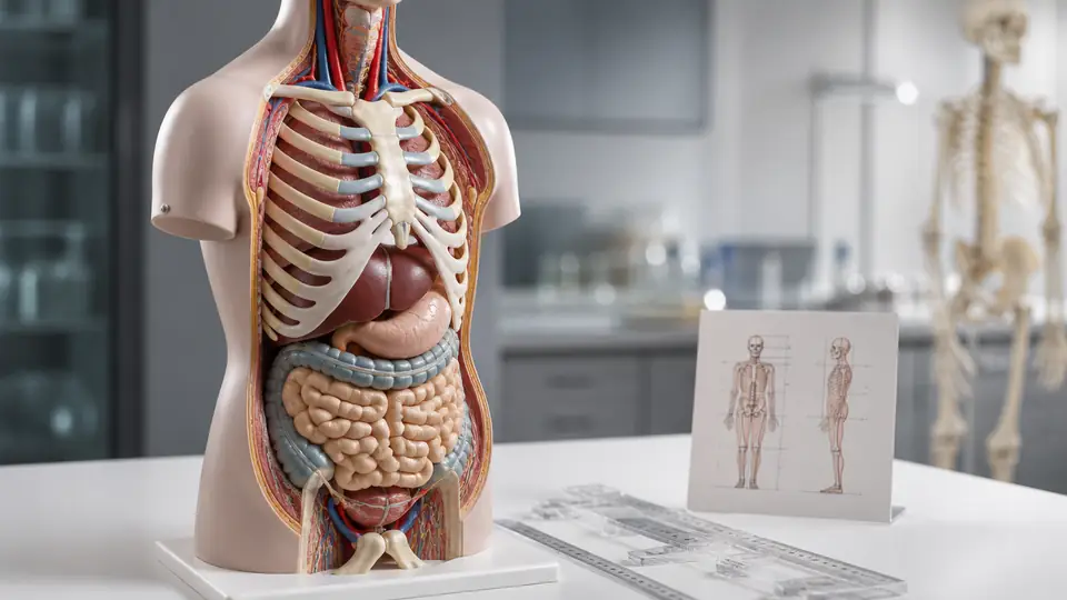
Stand beside a human anatomy model and the first impression is usually confidence: bones have familiar shapes, organs sit in recognizable regions, and colored vessels wind through the body like a tidy subway map. But look closer and a more useful question appears. How accurate are human anatomy models? The answer depends on what “accurate” means, what the model is designed to teach, and how closely its proportions and relationships match actual human anatomy.
An anatomy model is rarely a miniature human frozen in perfect detail. It is an educational representation. A classroom torso may enlarge small organs, separate structures so they can be seen, use bright colors that do not exist in the body, or leave out tissue layers that would hide the feature being taught. These are also useful distinctions to keep in mind when comparing life-size torso models, since “life-size” describes scale rather than guaranteeing that every structure has been reproduced exactly. Those choices can make the model more useful, but they also create limits that matter when a learner moves from a plastic display to an atlas, scan, cadaver, or clinical setting.
Accuracy Has More Than One Meaning
When someone asks how accurate a model is, they may be asking several different questions at once. Does it show the correct organs? Are those organs in the right places? Are the shapes, sizes, openings, attachments, and layers represented properly? A model may perform well in one category and only approximately in another. A simplified road map, for example, can preserve the relationships between important destinations without reproducing every bend, distance, or feature of the landscape.
For example, a heart model may accurately identify the four chambers, valves, major vessels, and direction of blood flow. It may still enlarge the heart, simplify the curvature of vessels, and exaggerate color differences between oxygenated and deoxygenated blood. A student studying valve location can benefit greatly from that design, while a learner trying to judge the exact external shape of a living heart needs a more cautious interpretation.
Accuracy is therefore best understood as fitness for purpose. A skeletal model intended to demonstrate joint movement has a different job from a detailed anatomical torso showing superficial muscles. A disarticulated skull designed for bone identification may separate parts that are tightly joined in an adult skull. The separation is not necessarily an error. It is a deliberate teaching feature, and the distinction becomes clearer when the model’s purpose is known.
What Anatomy Models Usually Represent Well
Major Structures and General Spatial Relationships
Well-designed models are often strongest at showing the broad arrangement of anatomy. A torso can place the lungs on either side of the mediastinum, position the heart between them, and show the liver beneath the diaphragm. A spine model can convey the alternating vertebral column, intervertebral discs, spinal canal, and broad curves of the cervical, thoracic, and lumbar regions. Those relationships are difficult to hold in the mind from a flat page alone.
That broad spatial picture is especially useful when a learner keeps confusing anterior with posterior or superior with inferior. Looking at the model from the front, then rotating it to the side, gives those directional terms physical context. The same student may still need an atlas for precise landmarks, but the model provides a three-dimensional framework into which those details can fit.
Large Bones, Organs, and External Landmarks
Large structures are usually easier to represent accurately than tiny or highly variable ones. The shaft and major ends of a femur, the lobes of the lungs, the general shape of the stomach, and the broad divisions of the brain can all be shown clearly at a useful scale. Enlarged models may even reveal grooves, chambers, or openings that would be too small to inspect on a life-size replica.
Consider a classroom skull. Its enlarged foramina and removable cranial bones may help a learner trace where nerves or vessels pass, even though the openings look more dramatic than they do in a real specimen. The model succeeds at making an important relationship visible. It should not be treated as a promise that every ridge and opening has been reproduced at exact scale.
Where Human Anatomy Models Simplify the Body
Scale and Proportion
Scale is one of the clearest sources of potential confusion. A model described as life-size is intended to approximate the dimensions of the represented anatomy, but even life-size models may differ because human bodies vary. A model listed as two or three times life-size is deliberately enlarged for visibility. Enlargement can improve a demonstration, yet it does not mean that every structure has been expanded with perfect mathematical consistency.
Imagine a teacher pointing to a model of the inner ear that is large enough for an entire class to see. The cochlea, semicircular canals, and vestibule become wonderfully legible, but their oversized presentation can distort a learner’s intuition about how small and delicate these structures are in the body. The model teaches organization and terminology very well. A separate reference is needed for actual dimensions.
Color Coding
Bright colors are usually instructional symbols, not literal anatomy. Red may indicate arteries, blue may indicate veins, and different shades may separate muscle groups, organs, or tissue layers. Human tissues do have natural color differences, but they do not generally appear as clean blocks of saturated red, blue, yellow, and purple.
This is easy to notice with a muscular figure whose muscles are painted in alternating colors. The design helps a student distinguish the biceps from the triceps or one abdominal muscle from another. It is less useful if the learner later assumes that the body contains those sharp color boundaries. A model’s legend, label key, or manufacturer documentation can clarify whether colors represent tissue, function, depth, or simply visual separation.
Omitted Tissue and Exposed Structures
Real anatomy is crowded. Fascia, connective tissue, fat, blood vessels, nerves, skin, and neighboring organs often overlap or conceal one another. Educational models remove some of that clutter so a particular structure can be seen. A torso may display the heart without covering it completely with the lungs, or show deep muscles that would normally sit beneath superficial layers.
That arrangement can create a useful teaching sequence. A learner might first study superficial muscles, then remove or set aside those pieces to examine deeper muscles. The model is not claiming that the body is naturally assembled with convenient handles and magnets. It is offering an artificial but informative way to explore depth, which is why model study works best alongside diagrams or sectional images.
Why Model Construction Affects Accuracy
Construction influences both what a model shows and how confidently it can show it. A rigid plastic model may preserve a stable shape and clear labels, while a flexible spine can demonstrate bending and disc movement. A sectional organ model may reveal internal cavities, but the cutaway surfaces are educational windows rather than normal anatomical surfaces.
Articulation introduces another layer of interpretation. A joint model with movable bones can demonstrate flexion, extension, rotation, or the relationship between adjacent vertebrae. When comparing functional joint models, however, it is worth remembering that visible movement is primarily an educational representation. A model may not reproduce the complete behavior of ligaments, cartilage, muscles, tendons, and soft-tissue restraints. When a knee model swings farther than a real knee would, the movement may be intended to show the direction of motion, not to establish a safe or normal range.
Removable parts create a similar tradeoff. A removable kidney or eye can be turned in the hand and examined from several angles, something a fixed model cannot offer. At the same time, repeated removal may require careful alignment, and the join between pieces may be thicker or more obvious than the natural boundary it represents. The number of detachable components can also be misleading unless you understand what part count means on a torso model. The educational gain is access, not perfect replication.
How Accurate Are Models for Different Learning Tasks?
| Learning Task | What A Model Usually Does Well | Where Caution Is Needed |
|---|---|---|
| Identifying major structures | Shows names, general shapes, and relative locations | Small landmarks may be simplified or omitted |
| Learning spatial relationships | Displays depth, orientation, and neighboring structures | Separated parts may not reflect natural overlap |
| Studying movement | Demonstrates broad joint directions and articulation | May not represent soft-tissue limits or normal ranges |
| Understanding layers | Allows superficial and deep structures to be viewed separately | Removed layers can make the body seem less crowded than it is |
| Learning fine detail | Enlargement can make selected features visible | Exact dimensions, texture, and variation may be inaccurate |
| Studying disease or injury | Can illustrate a specific structural change | A simplified depiction cannot represent every clinical presentation |
A student preparing for an introductory anatomy quiz may need a model that clearly separates the chambers of the heart or the bones of the foot. A medical educator discussing a surgical approach may need far more detail about tissue planes, vascular variation, and three-dimensional access. Both learners can use physical models, but the same model should not be assumed to satisfy both purposes equally well.
Even a highly detailed model usually represents typical anatomy rather than every natural variation. People differ in organ shape, vessel branching, bone contours, muscle attachments, and proportions. A model showing one arrangement can establish a useful reference point, but it cannot stand in for the entire range of human anatomy. That matters when a learner encounters a specimen, scan, or illustration that looks slightly different.
Physical Models Compared With Other Anatomy Resources
A physical model offers something a page cannot: the learner can view it from above, below, behind, and at an angle while maintaining a stable reference point. That is valuable for structures whose relationships are difficult to infer from separate two-dimensional images. A digital model may add rotation, transparency, sectional views, and labels that can be switched on and off, while a textbook may provide finer terminology, variation, and clinically relevant illustrations.
Neither format automatically wins. A digital heart may show blood flow or reveal chambers through a virtual cutaway, while a physical heart model makes scale and orientation tangible on a desk. A chart may label more structures in one view, while a removable model may help a learner understand which parts sit in front of others. Combining resources helps prevent any one representation from becoming mistaken for the body itself.
Cadaveric study, where available within an appropriate educational setting, provides another kind of information because it shows actual tissue relationships and variation. It also presents complexities that models intentionally reduce, including fragile tissues, overlapping structures, and changes caused by preservation. The contrast can be instructive. The clean model supplies an organized map, while real specimens show why anatomy is rarely as tidy as the illustration.
How to Judge the Accuracy of a Specific Model
Start with the intended educational level and subject. A general torso, a muscle figure, a vascular model, and a disarticulated skeleton are not judged by exactly the same standards. Check which structures are included, whether the model is life-size or enlarged, what parts are removable, and whether colors have a stated meaning. These details often reveal what the model is designed to communicate.
Next, compare the model with a dependable anatomy atlas, university teaching resource, or established anatomical reference. Look at relationships rather than counting tiny surface details first. Is the organ on the correct side? Are neighboring structures arranged in the correct order? Does the model distinguish the major regions accurately? A missing minor landmark may matter less in an introductory model than a misplaced major organ.
Labels deserve careful attention too. Some models use broad terms, while others identify smaller subdivisions. Terminology can also vary between educational resources, particularly where regions have common names and formal anatomical names. If a label seems unusual, verify it against an authoritative reference instead of assuming that the model or the textbook is automatically wrong.
Finally, treat manufacturer specifications as descriptions of that particular model, not as proof of universal anatomical truth. Claims about dimensions, scale, included pieces, and materials can be useful for understanding construction. They do not establish that the model reproduces every anatomical feature accurately. A model’s strongest evidence is the combination of clear design, appropriate purpose, and agreement with trusted anatomical references.
Common Misunderstandings About Anatomy Model Accuracy
“If It Looks Detailed, It Must Be Exact”
Fine texture, painted vessels, and numerous labels can create an impression of precision, but visual detail is not the same as anatomical fidelity. A model may include hundreds of visible structures while simplifying their depth, attachment, or proportions. Detail should be assessed feature by feature, not judged solely by how impressive the surface appears.
“A Model Shows the Only Normal Arrangement”
Most teaching models present a representative arrangement. They are not catalogs of every anatomical variation. A learner who sees an artery branch differently in another resource has not necessarily found an error. The difference may reflect normal variation, a different dissection plane, or a model that prioritizes another teaching objective.
“A Moving Model Reproduces Real Movement”
Movement in a model is usually selective. A hinged joint can show direction and broad mechanics, but real movement involves muscles, tendons, ligaments, cartilage, nerves, and changing contact surfaces. A flexible spine may illustrate curvature and segmental motion without representing the full behavior of living tissues. It is a demonstration of a relationship, not a complete biomechanical simulation.
Frequently Asked Questions
Are anatomy models actual size?
Some are designed to approximate life-size dimensions, while others are enlarged or reduced. Even a life-size model is an educational approximation because human anatomy varies and manufacturers may simplify proportions for visibility or construction. Always check the stated scale and dimensions for the specific model.
Are the colors on anatomy models realistic?
Usually, colors are primarily used for identification and separation. Red, blue, and other shades may indicate vessel types, tissue groups, or different anatomical structures rather than literal body coloration. A legend or labeling guide can explain the color system.
Can an anatomy model replace a textbook or atlas?
No single resource covers every aspect of anatomy. A model is particularly useful for shape, orientation, depth, and relationships. An atlas or textbook can provide more labels, terminology, sectional views, variation, and explanatory context. Using both gives a more complete picture.
Are expensive models always more accurate?
Price alone does not establish accuracy. Construction quality, scale, included structures, articulation, labeling, and intended audience all influence usefulness. A simpler model may be very effective for one lesson, while a highly detailed model may still simplify features that matter for another purpose.
The Practical Takeaway
Human anatomy models are generally accurate enough to teach major structures, spatial relationships, layers, and broad movement patterns, but they are not perfect replicas of individual human bodies. Their colors, scale, separable parts, exposed layers, and simplified surfaces are teaching decisions. The most reliable approach is to ask what the model is meant to show, verify important details with a trusted anatomical reference, and remember that one representation cannot capture every variation in real anatomy.
