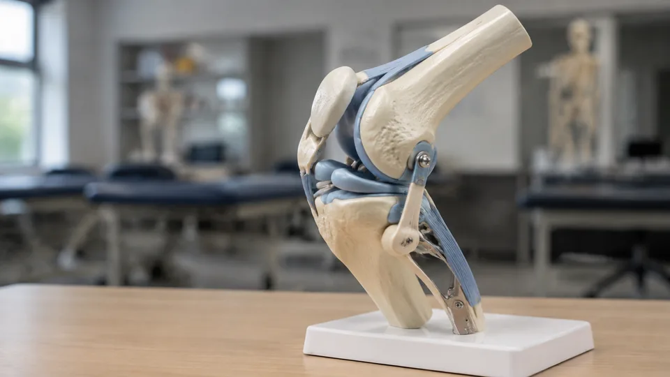
Twist a plastic shoulder and the difference becomes obvious within seconds. Some anatomy models glide through abduction and rotation while their flexible ligaments stay visible. Others barely move beyond a stiff hinge. Functional joint models vary sharply in anatomical detail, scale, soft-tissue representation, and the way they handle repeated classroom demonstrations.
Buyers often focus on bone shape while overlooking the structures that make a model genuinely useful. Flexible ligaments, visible cartilage, movable patellae, removable muscles, stable bases, and readable teaching guides can matter far more than a glossy finish. A physical therapy instructor may need a life-size knee for explaining ACL orientation, while a student working at a crowded desk may prefer a compact set covering several joint types.
For its balance of clear skeletal detail, functional motion, approachable size, and included photographic manual, the Axis Scientific Human Shoulder Joint Model is the strongest overall choice. The nine options below also cover premium knee study, removable hip muscles, multi-joint classrooms, and compact teaching setups.
Quick Answer
The Axis Scientific Human Shoulder Joint Model is the most versatile overall pick for demonstrating basic shoulder mechanics. Choose the 3B Scientific Deluxe Functional Knee Joint when realistic knee structures and premium educational support take priority. For a lab or classroom that needs broad coverage, the 6-Piece Human Joint Anatomy Model Set provides six life-size joint models rather than concentrating on one region.
Short on reading time? This table gives a quick view of the nine models and the practical differences that matter most.
Best Functional Joint Models Comparison Table
| Product | Best For | Scale or Coverage | Key Functional Feature |
|---|---|---|---|
| Axis Scientific Shoulder Joint Model | Overall shoulder education | Human shoulder | Flexible ligaments and multidirectional movement |
| AVASMODEL Functional Knee Model | Ligament identification | Life-size knee | Movable patella with major named ligaments |
| Hip Joint Model With Removable Muscles | Muscle relationships | Life-size, six parts | Magnetically attached removable muscles |
| Vision Scientific Functional Hip Model | Basic hip mechanics | Naturally sized hip | Flexible ligaments around a ball-and-socket joint |
| 3B Scientific Deluxe Knee Joint | Premium knee study | 12.6-inch-high model | Menisci, patella, ACL, PCL, and blue cartilage |
| 6-Piece Human Joint Anatomy Model Set | Classrooms and labs | Six life-size joints | Shoulder, elbow, hip, knee, hand, and foot coverage |
| Axis Scientific Life-Size Knee Model | Patient and sports education | Life-size knee | Ligaments, muscles, and gliding patella |
| Life-Size Muscled Shoulder Model | Rotator cuff study | 1:1 right shoulder | Multiple muscles, ligaments, and tendons |
| EVOTECH Mini Joint Model Set | Compact comparative study | Four half-scale joints | Portable shoulder, knee, hip, and elbow set |
The detailed reviews below show how each model fits a different study space, lesson plan, or demonstration style.
1. Axis Scientific Human Shoulder Joint Model | Best Overall

Shoulder anatomy becomes much easier to grasp once the humeral head can be moved against the shallow glenoid rather than viewed as two shapes on a page. This Axis Scientific model demonstrates abduction, adduction, and rotation while keeping its functional ligaments in view. Near-invisible attachment hardware helps the skeletal landmarks remain visually clear during a slow, close-range demonstration.
The model reproduces recognizable structures such as the humeral head, glenoid fossa, acromion, and coracoid process. That combination suits anatomy students, physical therapy instruction, chiropractic education, and calm one-to-one explanations across a clinic desk. Its full-color manual uses photographs of the actual model, so learners can move from a numbered image to the structure in their hands without mentally translating a different illustration.
Pros
- Demonstrates abduction, adduction, and shoulder rotation
- Functional ligaments remain visible as the joint moves
- Clear bone contours support three-dimensional landmark identification
- Photographic product manual corresponds directly to the model
- Backed by a three-year warranty and US-based support
Cons
- Focuses on skeletal and ligament anatomy rather than detailed shoulder musculature
- A single-joint model offers less comparative value than a multi-joint set
This is the sensible middle ground for buyers who want meaningful articulation without stepping up to a large muscled replica. It works particularly well when a learner needs to see why the shoulder gains mobility from its ball-and-socket arrangement, then trace the bony landmarks surrounding it. Those studying the rotator cuff muscle by muscle should look at the more elaborate shoulder model later in this list.
To buy this product, click here.
2. AVASMODEL Functional Life-Size Knee Model | Best for Ligament Study

Few joints create the same tangle of abbreviations as the knee. ACL, PCL, MCL, and LCL can blur together during early study, but this life-size AVASMODEL makes their relative positions tangible. The listing also identifies the posterolateral ligament and quadriceps tendon, giving learners more context than a stripped-down bone-and-hinge display.
Its movement includes flexion, extension, rotation, and patellar sliding. Picture an instructor bending the knee while a student watches the patella track along the front. That moving view is far more intuitive than memorizing isolated arrows. A removable base allows the model to alternate between shelf display and free handling, while the included color manual and downloadable resources provide useful prompts for guided study.
Pros
- Life-size format supports close inspection of knee structures
- Displays the ACL, PCL, MCL, LCL, and additional soft tissues
- Demonstrates flexion, extension, rotation, and patellar movement
- Provides 360-degree viewing for group or individual study
- Includes a removable base and supplementary teaching materials
Cons
- Dense ligament detail may feel busy for a very basic introductory lesson
- The listing does not specify individually removable ligament components
This knee is aimed squarely at students and instructors who want to point, bend, rotate, and compare ligament positions in one session. It is especially appealing for orthopedic anatomy and rehabilitation coursework where the patella and cruciate ligaments need to be discussed together. Buyers seeking more realistic bone coloration and clearly highlighted articular cartilage may prefer the premium 3B Scientific model.
To buy this product, click here.
3. Hip Joint Model With Removable Muscles, Six Parts | Best for Muscle Relationships

Peeling away a muscle layer can answer a spatial question that a crowded illustration leaves hanging. This life-size, six-part hip model uses removable muscles to reveal their relationship with the femur and hip bone. Muscle origins and insertions are represented, making the model useful for learners moving beyond the bare mechanics of a ball-and-socket joint.
Magnetic assembly adds a satisfying, practical rhythm to a lesson. A muscle can be removed, the underlying area discussed, and the piece returned without a jumble of tiny fasteners. The hand-painted PVC construction sits on a base and comes with a color educational brochure. It should suit nursing, anatomy, physiology, and movement-focused teaching where layers matter as much as articulation.
Pros
- Six-part life-size design provides layered anatomical study
- Removable muscles reveal underlying hip structures
- Shows muscle origins and insertions on the femur and hip bone
- Magnetic assembly makes repeated removal more convenient
- Mounted base and color brochure support display and teaching
Cons
- More assembly is involved than with a fixed ligament-only hip model
- Buyers focused purely on joint motion may not need the added muscle layers
This model makes the most sense for a learner who keeps asking, “What sits over that?” It offers a richer look at muscular relationships than the Vision Scientific hip, while the removable sections encourage active recall during a study session. For a quick demonstration of basic hip articulation with fewer pieces to track, the simpler Vision model remains the tidier choice.
To buy this product, click here.
4. Vision Scientific VAJ233 Functional Hip Joint Model | Best for Basic Hip Mechanics
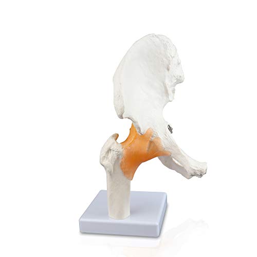
Clean, uncluttered anatomy has real teaching value. The Vision Scientific VAJ233 concentrates on the complete hip bone, a portion of the femur, and the surrounding flexible ligaments. With fewer colored muscle pieces competing for attention, the ball-and-socket relationship stays front and center as the joint articulates.
Its naturally sized format and mounted base fit a desk, demonstration counter, or study shelf without turning setup into a production. An instructor can pick up the model, show the femoral head sitting in the acetabulum, and move directly into permitted motion. This focused layout is particularly useful for introductory anatomy, where too much surface detail can feel like visual static.
Pros
- Naturally sized model gives the hip recognizable proportions
- Flexible ligaments help illustrate functional movement
- Complete hip bone provides useful pelvic context
- Uncluttered design keeps attention on joint mechanics
- Mounted base supports stable display between lessons
Cons
- Does not provide removable muscle layers
- Less suitable for studying detailed muscle origins and insertions
Choose this model for a straightforward explanation of how the hip articulates and how ligaments surround the joint. Its simplicity is a strength in short lessons, patient-facing conversations, or first-pass study. Advanced learners who need to lift muscles away and examine attachment areas will get more depth from the six-part removable-muscle model.
To buy this product, click here.
5. 3B Scientific A82/1 Deluxe Functional Knee Joint | Best Premium Knee Model
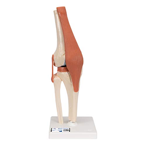
Realistic coloring gives this premium knee a striking presence on a teaching bench. Natural-cast bone tones sit beside blue-marked cartilage, making the articular surfaces pop during close inspection. The model includes portions of the femur, tibia, and fibula along with the menisci and patella, while the ACL and PCL are clearly defined.
At 12.6 inches high and mounted on a base, it is substantial enough for focused demonstrations without becoming cumbersome. Registration with 3B Smart Anatomy provides a warranty extension from three to five years and access to accompanying digital anatomy courses through the Complete Anatomy app. That physical-plus-digital pairing suits structured programs where a model on the table and an interactive view on a screen can reinforce the same lesson.
Pros
- Natural-cast bone coloration creates a realistic visual reference
- Blue cartilage clearly separates joint surfaces from bone
- Includes menisci, patella, ACL, and PCL
- Functional design is mounted on a stable base
- Eligible registration adds digital courses and a warranty extension
Cons
- Premium positioning may exceed the needs of casual home study
- Its single-joint focus offers less breadth than a classroom set
This is the pick for buyers who care about refined visual differentiation and a deeper educational package. The blue cartilage is particularly handy when a pale-on-pale model makes joint surfaces hard to distinguish under classroom lights. A beginner who needs a functional knee for occasional review can spend more conservatively, but a dedicated anatomy program may appreciate the added polish and digital support.
To buy this product, click here.
6. 6-Piece Human Joint Anatomy Model Set With Ligaments | Best for Classrooms

Lay six major joint models across a lab table and comparative anatomy suddenly becomes lively. This set covers the shoulder, elbow, hip, knee, hand, and foot at life size, with connected ligaments and functional articulation. Students can move from the broad rotation of a shoulder to the more constrained motion of an elbow without waiting for another model to circulate.
The PVC construction is described as lightweight and durable, a useful combination for teaching spaces where models regularly travel between shelves and workstations. Bending, rotation, and flexion vary by joint, so the set offers a broader biomechanics lesson than any single model here. It also gives physiotherapy, nursing, and anatomy classes a shared reference for both upper and lower limbs.
Pros
- Includes six distinct life-size joint models
- Covers shoulder, elbow, hip, knee, hand, and foot anatomy
- Connected ligaments support functional movement demonstrations
- Allows direct comparison of different joint mechanics
- Lightweight PVC construction is practical for repeated classroom handling
Cons
- Six models require considerably more storage and display space
- Individual joints provide less specialized soft-tissue detail than premium single-region models
A lab coordinator, tutor, or instructor teaching a full musculoskeletal unit will gain the most from this broad set. It is far easier to compare hand and foot organization when both are sitting side by side, ready to turn and inspect. Someone preparing for one knee examination or teaching a shoulder-specific module will probably get greater depth and less clutter from a dedicated model.
To buy this product, click here.
7. Axis Scientific Life-Size Knee Model With Ligaments and Muscles | Best for Demonstrations

A knee explanation often clicks at the exact moment the patella begins to glide. This Axis Scientific model demonstrates flexion, extension, rotation, and patellar movement while showing major ligaments, tendons, and muscle attachments. Internal hardware is concealed, keeping the viewer’s eye on anatomy rather than shiny screws cutting across the joint.
The included manual identifies 18 numbered structures using photographs of the model itself. That direct correspondence is helpful during self-study and quick demonstrations, since the learner can locate a number on the page and immediately hunt for it in three dimensions. A stable base keeps the model upright when it returns to the shelf, and the three-year warranty adds reassurance for settings where it may be handled often.
Pros
- Demonstrates several clinically relevant knee movements
- Movable patella makes tracking easier to visualize
- Includes visible ligaments, tendons, and muscle attachments
- Concealed internal hardware preserves a cleaner anatomical view
- Photographic manual labels 18 structures on the actual model
Cons
- Does not use highlighted blue cartilage like the deluxe 3B model
- Its detailed soft-tissue presentation may be more than a basic skeletal lesson requires
This model suits physical therapy classrooms, sports anatomy discussions, and one-to-one explanations where motion needs to remain visible from several angles. It offers more guided reference material than many basic knee models and keeps the moving patella at the heart of the demonstration. Buyers prioritizing highly differentiated cartilage and premium digital coursework should still consider the 3B option.
To buy this product, click here.
8. Life-Size Human Muscled Shoulder Joint Model | Best Detailed Shoulder

Where the top overall pick keeps the shoulder relatively clean, this 1:1 right-shoulder model embraces soft-tissue detail. It represents the infraspinatus, subscapularis, supraspinatus, teres major, and teres minor, along with the acromion, clavicle, humerus, scapula, and eight ligaments and tendons. Color differentiation helps prevent the dense cluster from turning into a beige anatomical knot.
The shoulder remains functional for demonstrating major movements, so the muscle coverage does not reduce it to a static sculpture. A removable base allows closer handling before the model is returned to display, and the PVC surface can be cleaned between teaching sessions. This is the sort of model that invites slower inspection, especially while tracing how the rotator cuff surrounds the humeral head.
Pros
- Full 1:1 scale supports detailed shoulder study
- Represents five named shoulder muscles
- Includes major bones plus eight ligaments and tendons
- Color-coded structures improve visual separation
- Functional movement and removable base support active demonstrations
Cons
- Dense soft-tissue detail can be overwhelming during introductory lessons
- Larger, more elaborate construction is less convenient for compact study spaces
This model belongs in a serious shoulder-focused setup where skeletal landmarks, rotator cuff muscles, and stabilizing tissues need to appear together. It makes sense for advanced students, educators, and practitioners conducting detailed educational conversations. For quick movement demonstrations or first-semester bone identification, the simpler Axis shoulder is easier to read at a glance.
To buy this product, click here.
9. EVOTECH Scientific Mini Joint Model Set | Best Compact Set

Desk space disappears quickly under notebooks, a laptop, and a life-size anatomical collection. EVOTECH’s half-scale set answers that problem with reduced-size shoulder, knee, hip, and elbow models. Each sits on a base and demonstrates relevant movement, giving students four major joint types in a format that remains manageable on a small table.
The listed heights range from roughly 4.75 inches for the shoulder to 7 inches for the knee, with the hip and elbow falling between them. Those compact dimensions make the set easy to reposition for tutoring, patient education, or a traveling presentation. The PVC material is also described as easy to clean, which suits models moving between hands during a busy practical session.
Pros
- Includes shoulder, knee, hip, and elbow models
- Half-scale construction saves desk and shelf space
- Separate bases keep all four models ready for display
- Supports comparison of several major joint movements
- Compact format is easier to carry between teaching locations
Cons
- Half-scale landmarks are harder to see from the back of a classroom
- Smaller structures offer less room for detailed close-up identification
This set is a smart fit for private tutors, students in small apartments, and educators who carry materials from room to room. Its charm lies in breadth without bulk. The trade-off is visibility, since a half-scale ligament cannot command a lecture hall like a life-size knee. For large groups, choose the six-piece life-size set or a dedicated full-size model.
To buy this product, click here.
How to Choose the Best Functional Joint Models
Match the Joint to the Lesson
Start with the anatomical question on the table. A shoulder model should make the shallow socket, humeral head, and surrounding stabilizers easy to see. A knee needs enough structure to distinguish the patella, menisci, collateral ligaments, and cruciate ligaments where provided. Buying a broad six-joint set for one focused ACL lesson creates clutter, while purchasing a lone hip for a full comparative anatomy course leaves obvious gaps.
Check What “Functional” Actually Means
Functional can describe anything from simple flexion at a hinge to multidirectional movement supported by flexible ligaments. Read the listed motions and visible structures rather than relying on that single word. For example, patellar sliding adds real educational value to a knee demonstration, while abduction and rotation are central to understanding the shoulder and hip.
Life Size Versus Compact Scale
Life-size models give fingers and eyes more room to identify small landmarks. They also read better when three or four students gather around a table. Half-scale sets trade that visibility for portability, a welcome compromise when the model must fit beside a laptop or travel in a teaching bag. In a large room, bigger usually communicates better.
Decide How Much Soft-Tissue Detail You Need
Flexible ligaments help explain stability and permitted movement, but muscles add another layer of complexity. Removable muscles are valuable for studying origins, insertions, and deeper relationships. Beginners may learn faster from a cleaner skeletal model where the acromion or acetabulum does not vanish beneath a crowded patchwork of colored tissue.
Look for Useful Color Coding and Guides
Color should clarify anatomy rather than decorate it. Blue articular cartilage, differentiated muscles, or numbered structures can speed up identification under bright study lamps. A guide featuring photographs of the exact model reduces friction because every angle and label matches the object sitting in front of the learner.
Consider the Base and Handling Style
A fixed base keeps a model upright and ready to display, which is handy in a clinic or classroom cabinet. Removable bases give learners more freedom to turn the joint over and inspect its underside. Multi-piece magnetic models encourage layer-by-layer exploration but require more care so detached components do not wander into a drawer.
Balance Breadth Against Depth
Multi-joint sets are excellent for comparing how an elbow differs from a shoulder or how a hand relates to a foot. Dedicated models usually offer more room for detailed ligaments, cartilage, muscles, or educational labeling. A classroom building a reusable collection may benefit from breadth first, followed by premium single-joint models for advanced modules.
Best Functional Joint Models FAQ
What Makes a Joint Model Functional?
A functional model articulates to demonstrate at least part of the joint’s natural movement. Flexible ligaments, movable bones, sliding patellae, or rotating ball-and-socket components may support that motion. The exact range varies by model, so the listed movements should match the lesson you plan to teach.
Are Life-Size Models Better Than Mini Models?
Life-size models are generally easier to inspect and show to a group. Mini models are lighter, more portable, and easier to store. A half-scale set works well for desk study or tutoring, while a life-size model is usually clearer for detailed landmark identification.
Should a Knee Model Include Both the ACL and PCL?
Both structures are useful for lessons covering internal knee stability and cruciate ligament orientation. Introductory bone study may not require that detail. Orthopedic, sports anatomy, and rehabilitation education typically benefit from clearly represented ACL and PCL structures.
Are Removable Muscles Worth Having?
Removable muscles are valuable when the course covers layered anatomy, attachment locations, and relationships between muscles and bones. They add setup time and visual complexity, so a ligament-only model may be more effective for quick demonstrations of basic joint mechanics.
Can Functional Joint Models Be Used for Patient Education?
Yes. A movable model can help illustrate general anatomy and show where a joint structure sits in relation to surrounding bones or ligaments. It should remain an educational visual aid rather than a substitute for individualized assessment, diagnosis, or professional medical instruction.
What Is the Most Useful Joint Model for Anatomy Students?
That depends on the course. A life-size knee with clearly visible ligaments suits detailed lower-limb study, while a functional shoulder explains multidirectional movement especially well. Students covering the full musculoskeletal system may get more mileage from a multi-joint set.
How Should an Anatomy Model Be Cleaned?
Follow the manufacturer’s care directions. Many PVC models can be wiped gently with a soft, slightly damp cloth. Avoid soaking flexible ligaments, painted surfaces, magnetic joints, or concealed hardware, and keep harsh cleaners away unless the maker specifically approves them.
Do More Parts Always Mean a Better Model?
No. Extra parts create more study layers, but they can slow a simple lesson and make a model harder to manage. The right level of detail is the level a learner can identify, explain, and use without the main joint mechanics disappearing under visual clutter.
Final Verdict
The Axis Scientific Human Shoulder Joint Model earns the top overall spot because it combines clear bony landmarks, functional ligaments, multidirectional movement, and a photographic manual in an approachable package. It is detailed enough for serious shoulder education yet clean enough that a beginner can follow the humeral head, glenoid, acromion, and coracoid without getting lost in layers of muscle.
For premium knee study, the 3B Scientific Deluxe Functional Knee Joint stands apart with realistic bone coloring, blue cartilage, menisci, cruciate ligaments, and digital learning support. The 6-Piece Human Joint Anatomy Model Set is the stronger classroom choice where broad comparison matters, while the compact EVOTECH Mini Joint Model Set fits tutors and students short on storage space. Match the joint, scale, and soft-tissue detail to the actual lesson, then favor clear movement over decorative complexity.
