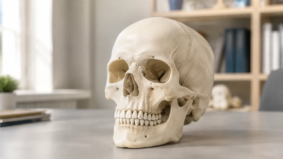
A flat atlas page can turn the skull into a maze of tiny labels. Pick up a three-dimensional model, remove the calvaria, trace the cranial base with a fingertip, and those same landmarks begin to make spatial sense. The best skull models for anatomy students vary sharply, though. Some emphasize numbered landmarks, others separate into individual colored bones, and a few include a removable brain for studying cranial relationships.
A common buying mistake is choosing a realistic-looking skull without checking how it supports study. Life-size proportions help with foramina and fossae, while color coding can make bone boundaries easier to memorize during a late-night review session. Numbered references, removable components, secure skull-cap connections, jaw articulation, and readable study materials often matter more than a dramatic bone finish. Priorities also differ. A medical student preparing for a practical may favor the JENACAD 55-landmark model, while an art student or commuter may prefer a compact exploded skull.
The JENACAD Human Skull Model is the strongest overall choice here because it combines life-size proportions, 55 numbered landmarks, painted sutures, three removable sections, and several study resources. The alternatives below cover simpler, more colorful, more portable, and brain-inclusive approaches.
Quick Answer
Best overall: The JENACAD Human Skull Model offers the most complete study system, pairing a numbered skull with contrasting sutures, a study card, a downloadable guide, and guided videos. Best color-coded option: The Skillssist Life-Size Colored Skull distinguishes 22 bones across nine colors and uses permanent laser-etched markings. Best skull-and-brain model: The QWORK 11-Part Model adds an eight-component brain, making it useful for visualizing how neural structures sit inside the cranial cavity.
For a faster scan before the detailed reviews, the table compares the nine selected models by format, removable parts, and defining study feature.
Best Skull Models for Anatomy Students Comparison Table
| Product | Best For | Format | Key Study Feature |
|---|---|---|---|
| JENACAD Human Skull Model | Overall study | Life-size, 3 parts | 55 landmarks, painted sutures, multimedia guides |
| Skillssist Colored Skull | Color-coded learning | Life-size, 3 parts | 22 bones in nine colors |
| QWORK Skull and Brain Model | Skull-brain relationships | Life-size, 11 parts | 3-part skull and 8-part brain |
| Doctor Verified 55-Landmark Skull | Cranial-base study | Life-size, 3 parts | Numbered foramina and magnetic cap |
| Laser-Etched Anatomical Skull | Frequent handling | Life-size, 3 parts | Permanent engraved markings |
| Classic Numbered Skull | Value-focused study | Life-size, 3 parts | 54 landmarks and painted sutures |
| Human Adult Skull Model | Uncluttered reference | Life-size, 3 parts | Realistic surface detail and reference chart |
| 15-Part Mini Exploded Skull | Portable bone study | Palm-sized, 15 parts | Individually colored detachable bones |
| EVOTECH Anatomy Model Set | Broad anatomy courses | Half-size skull plus body models | Skull, brain, skeleton, torso, and heart |
The reviews below take a closer look at how each model fits actual study sessions, lab preparation, teaching, and visual reference.
1. JENACAD Human Skull Model — Best Overall

This JENACAD model approaches the skull as an active-learning tool rather than a quiet shelf replica. Its 55 black numbered landmarks give students prompts for recall drills, while red-painted sutures stand out crisply against the bone-white PVC. Picture a practical exam looming on Friday night. Instead of hunting through an atlas for every structure, a student can identify a number, say the landmark aloud, and check the supplied study card or PDF.
The life-size model measures approximately 7.5 by 5.5 by 6.5 inches and weighs about two pounds, according to the listing. A magnetic calvaria and removable mandible divide it into three useful viewing sections. The spring-supported jaw moves for examining dentition, occlusion, and mandibular relationships, although gentle handling is sensible around its connectors. Guided study videos add another route for learners who absorb information better from demonstrations than dense pages.
Pros
- Features 55 numbered landmarks for structured recall practice
- Red-painted sutures make major cranial boundaries easy to locate
- Three-part construction exposes the calvaria, cranial base, and mandible
- Includes a printed study card, downloadable PDF, and guided videos
- Life-size proportions suit medical, dental, nursing, and A&P study
Cons
- Numbering and red sutures create a less natural-looking surface than an unmarked replica
- The spring-supported jaw requires careful pressure during repeated articulation
This is the most balanced option for students who want one skull to carry them from introductory bone identification into foramina, sutures, and practical-exam review. Artists seeking an uninterrupted bone-colored reference may prefer the plain model later in this list, but healthcare students who thrive on self-quizzing receive a far richer study setup here.
To buy this product, click here.
2. Skillssist Life-Size Colored Human Skull — Best Color-Coded Model

Color can turn a pale tangle of sutures into a much friendlier map. This Skillssist model represents 22 individual skull bones in nine vivid colors, helping visual learners separate neighboring structures at a glance. During a group review, one student can point to a colored region while another names the bone and its articulations. That quick visual feedback can be especially helpful early in an anatomy course, before every boundary feels familiar.
Laser-etched fonts and numbers complement the colored surface. Since the markings are engraved rather than handwritten, they are intended to remain legible through frequent handling and cleaning. The life-size PVC skull separates into the calvaria, skull base, and mandible, preserving the familiar three-part format used by many teaching models. It does not break all 22 colored bones into separate pieces, however. The coloring identifies boundaries visually while the model itself separates into three major components.
Pros
- Uses nine colors to distinguish 22 individual skull bones
- Laser-etched labels resist smudging during routine handling
- Life-size format supports realistic spatial study
- Three removable sections provide access to internal cranial surfaces
- Bright boundaries work well for visual learners and demonstrations
Cons
- Vivid colors are less realistic for students who want a natural bone appearance
- The 22 colored bones are identified visually rather than individually detachable
Choose this model if atlas color plates make more sense to you than rows of black numbers. It is particularly useful for beginning students, tutors, and instructors explaining bone boundaries across a table. Learners focused on subtle surface texture or realistic drawing reference may find the saturated palette distracting, but for fast identification, the visual clarity is hard to overlook.
To buy this product, click here.
3. QWORK Human Skull and Brain Anatomy Model — Best Skull-and-Brain Model

A skull alone explains the container. The QWORK model also shows what fits inside it, which gives neuroscience and broader anatomy students a more connected view. Its 11 components comprise a three-part skull and an eight-component brain. The brain includes cerebral hemispheres, a sagittal section, cerebellum, and brainstem, allowing a learner to lift sections away and inspect relationships that look frustratingly flat in a textbook diagram.
Both halves of the study system are numbered. The accompanying diagrams identify 55 markers on the skull and 32 on the brain, creating plenty of material for a partner quiz or classroom demonstration. Robust PVC keeps the set comparatively lightweight and easy to wipe clean. Since this model covers two complicated regions, individual skull details receive less singular emphasis than they do on a specialized exploded-bone model. Its real appeal is integration. You can move from the cranial cavity to the cerebellum and brainstem without swapping teaching aids.
Pros
- Combines a three-part skull with an eight-component brain
- Life-size format makes spatial relationships easier to visualize
- Includes diagrams for 55 skull markers and 32 brain markers
- Removable brain sections support sectional anatomy lessons
- Useful for neuroscience, patient education, and general anatomy classes
Cons
- More pieces require greater care during storage and transport
- Students studying individual cranial bones may prefer an exploded skull
This is the standout choice for anyone whose syllabus moves repeatedly between osteology and neuroanatomy. A nursing student reviewing cranial protection or an instructor showing brain placement gets more context from it than from a hollow skull. Students concentrating exclusively on the tiny features of individual facial bones can save space with a skull-only model, but the QWORK set offers broader teaching range.
To buy this product, click here.
4. Doctor Verified 55-Landmark Life-Size Skull — Best for Cranial-Base Study

Foramina at the cranial base can look like a jumble of pinholes and passages until a model lets you rotate them into view. This life-size skull numbers 55 major features, including landmarks around the skull base, so students can connect nerve pathways with physical openings. A downloadable PDF supplies the matching reference material. That setup suits a quiet desk drill: cover the key, identify each number, then rotate the skull and trace its position from another angle.
The model divides into the calvaria, skull base, and movable mandible. Strong embedded magnets hold the skull cap in place while retaining easy access to the endocranial cavity, and the listing emphasizes a tight cranial fit without conspicuous gaps. Detailed dentition and a smoothly opening jaw broaden its usefulness for dental anatomy and articulation study. High-density PVC provides a cleanable surface suited to repeated coursework, while the numbered finish keeps attention on identification rather than color-coded bone groups.
Pros
- Numbers 55 major landmarks, including skull-base features
- Strong magnetic connections secure the removable calvaria
- Movable mandible supports jaw and dental-structure study
- Life-size proportions provide a practical three-dimensional reference
- Downloadable PDF supports independent review away from class
Cons
- Lacks color-coded sutures or bones for learners who depend on visual grouping
- Three-part separation does not isolate individual cranial and facial bones
This model fits medical and dental students who already know the main bones and need to sharpen landmark recognition, particularly around the cranial base. Its secure magnetic cap is useful when a model gets passed around a study group. Beginners who still confuse adjacent bones may progress faster with the colored Skillssist model, while advanced visual-spatial review is the stronger suit here.
To buy this product, click here.
5. Upgraded Laser-Etched Anatomical Skull — Best for Frequent Handling

Handwritten markings can become grubby or faint after countless fingers circle the same suture. This upgraded model addresses that annoyance with laser-etched fonts and numbers engraved into the surface. The lettering is uniform, permanent, and intended to stay readable through touch and routine cleaning. For a tutoring lab where the skull travels from hand to hand every week, that durability has genuine practical value.
Beneath the engraved finish is a conventional life-size, three-part teaching skull. Students can remove the calvaria, inspect the base, and articulate the mandible while following the included instruction sheet. The non-toxic PVC construction is tasteless and easy to maintain. Its presentation is comparatively straightforward. There are no separate brain sections, multicolored bones, or extensive multimedia lessons competing for attention. That simplicity works for learners who want labels fixed directly beside the anatomy, though it offers less guided support than the top-ranked model.
Pros
- Laser-etched fonts and numbers resist fading and smudging
- Uniform engraving is easier to read than irregular handwritten labels
- Separates into the calvaria, skull base, and mandible
- Life-size format works for individual study and teaching tables
- Cleanable PVC construction suits repeated handling
Cons
- Included instructional support is simpler than the multimedia resources supplied with some rivals
- Monochrome presentation offers less visual separation between neighboring bones
Teachers, tutors, and study groups should give this model a close look if marking longevity carries more weight than colorful presentation. The engraved surface removes anxiety about labels rubbing away during a semester of drills. A solo beginner may get more direction from a model with videos or a detailed key, but busy shared spaces can benefit from this model’s sturdy, uncluttered approach.
To buy this product, click here.
6. Classic Numbered Human Skull Model — Best Value Numbered Model
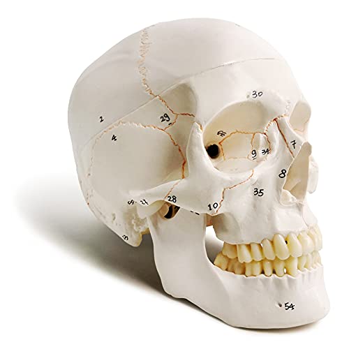
This classic model packages the core features many college students need without drifting into an elaborate kit. It stands nine inches tall, follows life-size adult proportions, and carries 54 numbered landmarks in permanent ink. Hand-painted suture lines provide an immediate visual route across the cranium. During a rapid-fire study session, those lines make it easier to follow the coronal, sagittal, lambdoid, and squamous regions before checking finer landmarks against the supplied chart.
The skull separates into three sections and uses small magnets to keep the cranial opening secure. That prevents the cap from sliding away as the model is tilted to expose inferior structures. A spring secures the lower jaw while still allowing articulation, and the package includes a manual plus study chart. The listing states that the model was cast from a real human specimen, which contributes to its realistic proportions and surface reference. Painted lines and permanent-ink numbers remain less durable than deep engraving, so sensible cleaning is appropriate.
Pros
- Includes 54 numbered bone landmarks and a matching reference sheet
- Painted sutures make major cranial boundaries more visible
- Nine-inch life-size format supports practical anatomy study
- Magnetic cranial cap stays secure while the model is tilted
- Articulated spring-held mandible allows basic jaw demonstration
Cons
- Permanent-ink numbers are less robust than laser-etched markings
- Hand-painted sutures may appear less uniform than molded or engraved boundaries
Students seeking a practical balance between labeling, life-size scale, and supporting materials will find plenty to work with here. It is well suited to an introductory college anatomy desk, where a student wants to quiz landmarks and then look inside the cranial vault. Shared laboratories expecting heavy cleaning may favor the engraved model above, but this classic option offers a useful feature set for personal study.
To buy this product, click here.
7. Human Adult Skull Anatomical Model — Best Uncluttered Reference

Labels are handy until the goal is to recognize anatomy without a prompt. This nine-inch adult skull keeps the surface visually clean, letting fissures, processes, sutures, and contours do the talking. The listing says it was cast from a real adult specimen, giving students and artists a realistic structural reference. Imagine sketching the orbit under a desk lamp or practicing a no-hints bone identification drill. The absence of numbers forces attention back onto shape and location.
Like many full-size educational skulls, it separates into the calvaria, base, and mandible. Magnets and small pegs secure the removable cap, while the articulated lower jaw allows basic movement and positional study. Non-toxic PVC is washable, tasteless, and suited to routine classroom use. A colorful skull chart supplies labels separately, so students can consult guidance without permanently covering the model in visual cues. This separation between model and key is simple, but pedagogically useful for closed-book recall.
Pros
- Clean, unnumbered surface supports no-prompt identification practice
- Life-size nine-inch format provides realistic proportions
- Three-part design reveals internal and external cranial structures
- Magnetic cap uses small pegs for more secure alignment
- Includes a colorful reference chart for guided study
Cons
- Finding small structures takes longer without numbers printed on the model
- No color coding separates adjacent cranial and facial bones
Artists, educators, and students who have moved beyond basic label matching should appreciate this skull’s quiet, natural presentation. It also works well for mock practicals because nothing printed beside a landmark gives the answer away. A nervous first-semester learner may prefer numbered cues, but anyone building true visual recognition can use the separate chart only when needed.
To buy this product, click here.
8. 15-Part Mini Exploded Skull Model — Best Compact Option
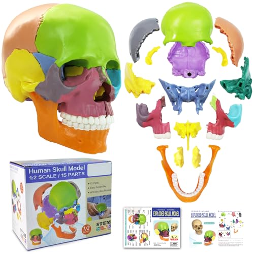
The palm-sized format makes this colorful skull easy to carry from a dorm desk to a library study room. More importantly, it separates into 15 components rather than three broad sections. Paired maxillae, cheekbones, nasal bones, lacrimal bones, palatine bones, and turbinate bones join the vomer, mandible, hyoid, and other cranial components. Pulling these pieces apart can make the orbit and nasal cavity feel far less mysterious than they do on a sealed model.
Different colors distinguish the bones and paired structures, while discreet connectors along simplified sutures support reassembly. The surface includes eye sockets, canals, cavities, teeth, and visible texture in a compact package. An illustrated guide adds general information and review questions. Portability is the headline benefit, but scale creates a clear compromise. Tiny openings are harder to inspect than on a life-size skull, and a palm-sized model will not command a large classroom from the back row.
Pros
- Separates into 15 components for studying individual bones
- Color coding makes paired and neighboring bones easier to distinguish
- Palm-sized body travels easily in a backpack or study bag
- Shows internal nasal, orbital, and endocranial relationships
- Includes an illustrated guide with study questions
Cons
- Small scale makes fine foramina and surface landmarks harder to inspect
- Numerous compact pieces require careful storage to avoid loss
This is a clever companion for students who learn by taking structures apart and rebuilding them. It is also appealing to art and cosmetic-medicine learners exploring how facial bones form the orbit, nose, and mouth. It should not be the first choice for detailed cranial-base practicals, but as a colorful spatial puzzle that fits beside a laptop, it earns its place.
To buy this product, click here.
9. EVOTECH Human Body Anatomy Model Set — Best for Broad Anatomy Courses

Some courses race from bones to organs in a matter of weeks, leaving little reason to buy a large model for every unit. The EVOTECH set addresses that broader curriculum with a half-size skull and brain alongside skeleton, torso, and heart models. Its skull is about 3.5 inches, while the skeleton stands roughly 17.7 inches, the torso 11 inches, and the heart about eight inches including its base. Everything packs into an included carrying case.
The components are hand-painted and come with separate guides, including a skull-and-brain manual, skeleton chart, heart manual, and human-body brochure. A student can review cranial anatomy, shift to the axial skeleton, and finish with cardiac structures using one compact collection. The trade-off is depth. The small skull cannot match the visibility of a life-size 55-landmark model, and much of the package is devoted to anatomy beyond the head. Still, for homeschooling, introductory survey classes, and mobile teaching, that breadth is precisely the point.
Pros
- Includes skull, brain, skeleton, torso, and heart models
- Broad coverage suits introductory and whole-body anatomy courses
- Comes with multiple illustrated manuals and reference materials
- Included carrying case simplifies transport and storage
- Compact models can fit into small study or teaching spaces
Cons
- Half-size skull is less suitable for studying very small cranial landmarks
- Students focused solely on skull anatomy pay for several unrelated body models
This set makes the most sense for instructors, families, or early-stage students who need a portable tour of human anatomy rather than maximum cranial detail. A learner preparing for a skull-specific laboratory practical should choose one of the life-size options above. For a survey course where tomorrow’s lesson jumps from the brain to the heart, the EVOTECH collection offers useful range in one case.
To buy this product, click here.
How to Choose a Skull Model for Anatomy Study
Start With the Course Objective
Match the model to the next task on the syllabus. Identifying cranial bones requires a different tool from tracing nerves through skull-base foramina. A first-semester student staring at unfamiliar sutures may benefit from nine bright colors, while a medical student preparing for a no-label practical needs realistic proportions and fewer visual hints. Look at your laboratory checklist, then choose the model that turns those exact structures into something visible and testable.
Brain-inclusive models have a separate advantage. They help explain how the cerebrum, cerebellum, and brainstem occupy the cranial cavity, which is useful in neuroscience and broad anatomy instruction. Their extra components bring more storage and assembly, so a skull-only model remains cleaner for dedicated osteology work.
Life-Size Versus Compact Models
Life-size skulls give small foramina, processes, fossae, and dental structures enough room to inspect from several angles. They also translate more naturally to practical exams that use full-size specimens or models. The cost is bulk. A large skull occupies precious desk space and can feel awkward inside a crowded backpack.
Compact models make excellent secondary study aids. A palm-sized exploded skull can sit beside lecture notes at a coffee shop, and separating individual colored bones creates a memorable little puzzle. Fine details inevitably become smaller, however. For deep cranial-base work, a portable miniature is better viewed as a supplement than a replacement for life-size anatomy.
Numbered, Colored, or Unmarked
Numbered landmarks create a ready-made quiz. Cover the reference key, call out the structure, and check the answer. This method keeps a review session moving, especially when a study partner is firing questions across the table. Confirm that the matching key is included or available digitally, since bare numbers have little educational value without a dependable reference.
Color-coded bones provide faster boundary recognition. They are particularly helpful while learning which bones form the orbit, nasal cavity, and cranial vault. An unmarked bone-colored skull is harder at first, but it removes cues and looks closer to the neutral models often used for formal identification. Students sometimes pair approaches: learn boundaries on color, then practice recall on a plain model.
Removable Parts and Connections
A three-part skull typically separates into the calvaria, base, and mandible. That configuration is sufficient for examining the cranial cavity and jaw while keeping setup straightforward. Magnets make the cap quick to remove, and alignment pegs help it settle back into position without an annoying scrape or wobble.
Exploded skulls go much further by separating individual cranial and facial components. They reveal how delicate pieces contribute to the orbit, palate, and nasal architecture. More pieces mean more opportunities for loss, so use a divided storage container and count every component after a group session. Tiny connectors also deserve patient handling rather than force.
Jaw Articulation
An articulated mandible helps students visualize dental position, occlusion, and the relationship between the jaw and cranium. Spring-supported jaws are common on educational models and allow basic opening motion, but they do not reproduce the full complexity of living temporomandibular movement. Use them for structural demonstration rather than biomechanical simulation.
Check how the mandible detaches if your coursework requires close inspection of its ramus, body, condyle, or foramina. A removable jaw is easier to rotate in the hand, while a firmly mounted one can be more convenient during quick classroom demonstrations.
Marking Durability and Cleaning
Laser-etched labels offer an advantage in shared laboratories because the markings are physically engraved. Permanent ink is perfectly practical for personal study, although aggressive scrubbing may be less appropriate. Painted sutures add clarity but also call for gentle cleaning. A soft damp cloth is usually a safer approach than harsh chemicals unless the manufacturer provides specific care directions.
PVC appears throughout this category because it is lightweight and easy to maintain. Build quality still varies by model design, particularly around magnets, pegs, springs, and small detachable bones. Store the skull with its cap properly seated and avoid lifting it by the mandible, where leverage can strain the connectors.
Study Materials and Teaching Support
A model is much more useful when its numbers match a readable chart, PDF, or manual. Digital files travel well to the library, while printed cards are faster during a tabletop quiz. Videos can help students learn how to orient the skull before memorizing dozens of landmarks. Confirm what is supplied in the listing, since the depth of study support differs widely.
Reference materials should complement an accredited course atlas and instructor guidance. A commercial skull model is an educational visualization aid. It does not replace cadaveric study, laboratory instruction, clinical training, or authoritative anatomical references.
Best Skull Models for Anatomy Students FAQ
Is a Life-Size Skull Better for Anatomy Students?
A life-size skull is usually better for inspecting small foramina, fossae, processes, sutures, and tooth relationships. It also provides a more realistic sense of proportion. Compact skulls remain valuable for portability and bone-assembly practice, especially when they separate into many colored components.
Should I Choose a Numbered or Unnumbered Skull?
Choose a numbered model for guided self-testing and quick reference. An unnumbered model is useful for mock practicals, drawing, and recall without printed hints. Students who can afford the space sometimes learn on a numbered or colored skull, then test themselves on a plain one.
How Many Parts Should an Anatomy Skull Have?
Three parts are enough for most general study because the removable calvaria exposes the cranial cavity and the detachable mandible allows separate inspection. A 15-part exploded model is more useful for understanding how individual facial and cranial bones fit together. Skull-and-brain sets add value for neuroanatomical relationships.
Are Color-Coded Skull Models Anatomically Useful?
Yes. Color coding makes adjacent bones and major boundaries easier to distinguish, which can reduce confusion during early coursework. The colors are educational rather than realistic, so advanced students should still practice recognizing structures by shape, location, and articulation.
Do Medical Students Need a Model With Numbered Foramina?
Numbered foramina can make cranial-base review faster by linking each opening to a reference key. They are useful for studying pathways and preparing for identification exercises. Students should still learn each landmark’s position and relationships rather than memorizing the printed number alone.
Is a Skull-and-Brain Model Better Than a Skull Alone?
It depends on the course. A skull-and-brain model is stronger for visualizing how major brain regions sit within the cranial cavity. A skull-only model generally provides a simpler setup for osteology, sutures, foramina, and facial-bone study.
How Should an Anatomical Skull Model Be Cleaned?
Follow the manufacturer’s care guidance. For routine dust and fingerprints, gentle wiping with a soft, lightly damp cloth is a cautious starting point. Avoid soaking magnetic or spring-connected sections, and keep harsh cleaners away from painted or inked markings unless the listing confirms compatibility.
Can a Skull Model Replace an Anatomy Atlas?
No. A model provides three-dimensional orientation, while a good atlas supplies standardized labels, sectional views, and deeper contextual information. The two work best together. Use the model to trace structures physically, then verify names and relationships with course-approved references.
Final Verdict
The JENACAD Human Skull Model is the strongest overall recommendation among these nine picks. Its life-size format, 55 landmarks, sharply painted sutures, removable calvaria and mandible, articulated jaw, study card, PDF, and guided videos create a coherent system for active recall. It is especially well matched to medical, nursing, dental, PA, therapy, and anatomy students preparing for practical identification rather than buying a skull as passive desk decor.
Different study habits point toward worthwhile alternatives. The Skillssist Life-Size Colored Skull gives visual learners exceptionally clear bone separation, while the QWORK Skull and Brain Model adds valuable context for neuroanatomy. Budget-conscious students wanting a traditional numbered format can look to the Classic Numbered Human Skull, and commuters may prefer the portable 15-Part Mini Exploded Skull. Before choosing, compare your syllabus with the model’s scale, markings, removable sections, and reference materials. The right skull should make your next study session more active, more spatial, and far less like squinting at a crowded page.
