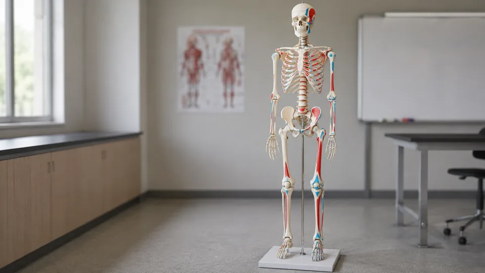
A flat anatomy chart can make muscle attachments feel like a maze of red and blue labels. Put a marked skeleton beside it, though, and the picture gains depth. You can trace an attachment around a humerus, rotate a foot to visualize movement, or point out landmarks to a class without squinting at a page. The best skeleton models with muscle markings make those connections clear while remaining sturdy enough for repeated study and demonstration.
Models differ sharply in scale, articulation, labeling, and added structures. A life-size skeleton on casters works beautifully in a classroom or clinic, yet it can overwhelm a cramped study corner. A 31-inch model fits on a desk, while focused hand and foot models reveal regional anatomy more clearly. Buyers often focus on size alone and overlook the quality of the muscle-origin and insertion markings, stand stability, removable parts, or joint movement. The 70.8-inch Human Anatomy Model earns our top overall spot by combining full-body muscle markings, numbered bones, ligaments, nervous-system details, and practical storage features.
For a faster decision, the quick picks and comparison table highlight the strongest choices before the full reviews.
Quick Answer
Best overall: The 70.8-Inch Human Anatomy Model offers a broad mix of muscle attachment markings, numbered bones, flexible joint ligaments, removable cranial components, and a rolling stand. Students short on floor space should look at the Axis Scientific Mini Human Skeleton. For concentrated regional study, the EVOTECH hand and Merinden foot models provide life-size anatomy without requiring room for a complete skeleton.
Best Skeleton Models With Muscle Markings Comparison Table
| Product | Best For | Scale or Size | Key Anatomical Features |
|---|---|---|---|
| 70.8-Inch Human Anatomy Model | Best overall | 70.8 inches, life-size | Muscle markings, numbered bones, ligaments, nerves, carotid artery |
| breesky Painted Skeleton Model | Classroom teaching | 70.8 inches, life-size | Painted attachment points, numbered bones, removable parts |
| Life-Size Skeleton With Spinal Nerves | Joint demonstrations | 71 inches, life-size | Spinal nerves, joint ligaments, muscle origins and insertions |
| breesky Skeleton With Nervous System | Layered anatomical reference | 70.8 inches, life-size | Muscle markings, ligaments, nervous system, numbered bones |
| Axis Scientific Mini Skeleton | Compact study | 31 inches | Painted and numbered attachment points, articulated joints |
| EVOTECH Hand Skeleton | Hand anatomy | Life-size right hand | Painted origins and insertions, articulated fingers and wrist |
| Merinden Foot and Ankle Model | Foot biomechanics | Life-size right foot | Red attachment markings, articulated joints, tibia and fibula |
| Original 180 cm Skeleton Model | Premium institutional display | 180 cm, life-size | Enumerated bones and muscles, replaceable teeth, stand |
| Nine-Part Foot Skeleton Model | Detailed foot dissection | Life-size right foot | Muscles, ligaments, nerves, vessels, removable layers |
The detailed reviews below explain where each model fits, from lecture rooms and clinics to crowded student desks.
1. 70.8-Inch Human Anatomy Model: Best Overall
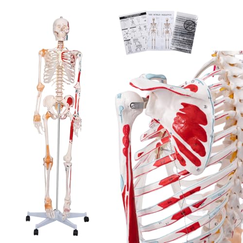
This model earns first place by covering the anatomical features most buyers seek in one life-size teaching aid. Painted muscle origins and insertion points turn the pale PVC bones into a readable attachment map, while numbered bones support identification exercises. Flexible joint ligaments and visible nervous-system and carotid-artery structures add useful context for lessons that extend beyond memorizing individual bones.
The practical details matter once a skeleton becomes part of a busy room. Its five-wheel metal stand lets an instructor roll it from a storage corner to the front of a class, and the supplied dust cover protects all those painted surfaces between sessions. The skull cap comes off, the jaw moves, and three teeth are detachable. Hinged limbs and joints provide posing flexibility for demonstrations, although buyers should expect educational approximation rather than natural human biomechanics.
Pros
- Life-size format supports room-wide teaching and realistic landmark comparison
- Shows muscle origins, insertions, numbered bones, joint ligaments, and neural details
- Removable skull cap, movable jaw, and detachable teeth expand cranial study
- Five-wheel metal stand makes repositioning easier
- Includes a dust cover, color illustrations, assembly instructions, and a poster
Cons
- Requires substantially more floor and storage space than a miniature model
- Its extensive detail may be more than beginners need for basic bone identification
This is the strongest choice for an anatomy classroom, physical therapy office, or teaching space that needs one adaptable full-body reference. Its blend of markings and added structures offers more lesson flexibility than a bare-bones model. Apartment-based students and occasional hobby learners may prefer the smaller Axis model, but regular instructors can make good use of this model’s broad anatomical coverage.
To buy this product, click here.
2. breesky 70.8-Inch Painted Skeleton Model: Best for Classrooms

Picture a lecturer moving from the shoulder girdle to the lower leg while students follow the colored attachment points from several rows away. The breesky model is built for that familiar classroom rhythm. It stands 70.8 inches tall, has painted muscle-origin and insertion areas, and includes numbered bones, giving instructors several visual reference systems within one model.
The skull, arms, and legs are detachable, which can make small-group demonstrations easier. Rather than crowding around the entire skeleton, a student can work with an individual limb or examine the jaw and teeth through the removable skull. Three educational posters broaden the package for wall-based reference. Its PVC construction can be wiped clean, and a five-wheel stand helps it travel between a laboratory bench, lecture area, and office without awkward lifting.
Pros
- Full-size proportions make major landmarks easy to show to groups
- Combines painted muscle attachment areas with numbered bones
- Detachable arms, legs, and skull support focused demonstrations
- Five-wheel stand is convenient for shared classrooms and labs
- Includes three educational posters for complementary visual study
Cons
- The spine is fixed rather than flexible
- Large footprint is poorly suited to a small bedroom or crowded desk area
Choose this breesky skeleton if straightforward classroom usability matters more than an unusually dense set of nerves and vessels. The detachable limbs are particularly handy for practical sessions where several learners need a closer view. Buyers hoping to demonstrate spinal flexion should skip it because the listing expressly identifies the spine as nonflexible.
To buy this product, click here.
3. Life-Size Skeleton Model With Spinal Nerves: Best for Joint Demonstrations

For lessons where a static pose tells only half the story, this 71-inch model brings articulation into focus. The listing identifies movement at the shoulder, lower jaw, elbow, wrist, knee, and foot, creating opportunities to connect painted muscle origins and insertions with joint position. Joint ligaments and spinal nerves add another visual layer for nursing, physiology, and musculoskeletal instruction.
The removable calvarium opens the cranial area for closer examination, while detachable arms and legs can simplify regional teaching. Its hand-painted PVC construction is intended to provide a realistic three-dimensional reference and remain easy to clean. A detachable rolling stand with four casters supports transport, a practical benefit when a model spends the morning in a classroom and the afternoon helping explain anatomy in an office.
Pros
- Life-size 71-inch build is suitable for full-body instruction
- Marks muscle origins and insertion points
- Includes spinal nerves and joint ligaments for added context
- Articulation across several major joints supports movement demonstrations
- Removable calvarium and detachable stand increase teaching flexibility
Cons
- Four-caster stand offers one fewer contact point than the five-wheel stands listed for several rivals
- Full-size dimensions demand a dedicated storage location
This model makes sense for educators who regularly connect bones, muscle attachments, nerves, and joint movement in the same lesson. It is especially appealing for demonstrations around the shoulder, elbow, knee, and foot. Someone seeking a compact revision tool will get easier day-to-day handling from the Axis Scientific model, while this option belongs where a large visual reference can stay assembled.
To buy this product, click here.
4. breesky Skeleton Model With Nervous System: Best Layered Reference

A basic skeleton answers “where is the bone?” This 70.8-inch breesky model supports the next set of questions by combining muscle origins and insertions with flexible joint ligaments, numbered bones, and nervous-system details. That layered presentation can help learners visualize how separate structures occupy the same body rather than studying each system on an isolated chart.
The removable skull permits closer inspection of the jaw and teeth, while detachable limbs make the model easier to use for regional instruction. Imagine a small lab group carrying an arm to a table and tracing attachments around the elbow instead of circling a tall stand. The five-wheel base handles moves across smooth classroom floors, and three included posters provide quick reference when the physical model is being used by another group.
Pros
- Combines muscle markings, ligaments, nerves, and numbered bones
- Life-size proportions work well for lectures and patient education
- Removable skull and detachable limbs enable close regional viewing
- Five-wheel rolling stand supports movement between teaching areas
- Includes three posters to reinforce visual identification
Cons
- Nonflexible spine limits demonstrations of spinal movement
- Dense visual information may feel busy during introductory bone lessons
This is a sensible pick for multidisciplinary teaching where instructors move between skeletal, muscular, ligamentous, and neural structures. Its advantage over the simpler breesky painted skeleton is the added nervous-system context. For an uncluttered osteology course, the extra layers may distract from the immediate lesson, so the intended curriculum should guide the choice.
To buy this product, click here.
5. Axis Scientific Mini Human Skeleton Model: Best Compact Option

A life-size skeleton can dominate a room like an extra roommate. The 31-inch Axis Scientific model is a friendlier proposition for a dorm desk, home office, or compact tutoring space. It retains a full-body view and uses painted, numbered muscle insertion and origin points, allowing students to move between a textbook illustration and a three-dimensional reference without finding floor space for a six-foot model.
Articulation is represented at the shoulders, elbows, wrists, hips, knees, and ankles. The removable white base and metal stand keep the model upright while leaving the miniature skeleton accessible for study. Axis also supplies a full-color product manual identifying model features, which is valuable when a painted patch makes sense visually but its name refuses to spring to mind during revision.
Pros
- Compact 31-inch height fits desks, shelves, and small study rooms
- Painted and numbered muscle origins and insertions aid identification
- Articulated major joints add movement context
- Removable base provides display flexibility
- Includes a full-color identification manual
Cons
- Reduced scale makes fine landmarks harder to inspect than on life-size models
- Smaller presence is less effective for teaching a large room
Students, artists, tutors, and home learners are the natural audience here. It offers full-body orientation without the storage headache or visual bulk of a life-size skeleton. A lecturer addressing a packed classroom should choose a larger model, but one person studying at arm’s length may find this miniature far easier to live with and revisit daily.
To buy this product, click here.
6. EVOTECH SCIENTIFIC Hand Skeleton Model: Best for Hand Anatomy

The hand packs many small bones and attachment sites into a remarkably tight area. A full skeleton can make those structures feel tiny and fiddly, while this life-size right-hand model places the carpals, metacarpals, phalanges, wrist, and lower portions of the radius and ulna at center stage. Painted muscle insertions and origins help connect bony landmarks with the forces that move the fingers and wrist.
Stainless wire provides articulation, so learners can manipulate the hand and explore relationships during movement. That tactile element can be far more memorable than staring at a rigid plastic pose before an exam. The PVC model sits on a sturdy ABS base for display and comes with an instruction guide, making it appropriate for an anatomy classroom, practitioner’s office, art studio, or student desk.
Pros
- Life-size format preserves realistic hand and wrist proportions
- Painted origins and insertions clarify muscular attachment areas
- Articulated wire construction allows finger and wrist movement
- Includes portions of the radius and ulna for forearm context
- Stable display base and instruction guide suit regular study
Cons
- Regional format cannot teach whole-body skeletal relationships
- Wire articulation approximates movement rather than reproducing every natural joint constraint
This focused model is a sharp choice for learners who need more hand detail than a full-size skeleton can comfortably show. It also suits artists studying finger poses and educators explaining basic wrist mechanics. Buyers building a general anatomy resource should begin with a whole skeleton, then add this model when upper-limb coursework demands a closer look.
To buy this product, click here.
7. Merinden Painted Foot and Ankle Anatomy Model: Best for Foot Biomechanics

Foot anatomy becomes much clearer once the model can be turned, tilted, and viewed from beneath. This life-size right-foot skeleton includes the ankle, tibia, and fibula, preserving the lower-leg context that helps explain joint stability. Red muscle-origin and insertion markings make attachment areas visible across the bones without covering the skeletal shapes under layers of molded muscle.
The articulated design supports demonstrations of foot and ankle movement, gait concepts, and basic range-of-motion relationships. A stable base keeps the model upright between lessons, sparing delicate joints from rolling around a crowded desk. For a podiatry student puzzling over a flat diagram or an educator explaining why the ankle involves more than a simple hinge, the three-dimensional view can make the lesson click.
Pros
- Life-size proportions support realistic regional comparison
- Red muscle attachment markings are easy to distinguish from bone
- Articulated joints help demonstrate foot and ankle movement
- Included tibia and fibula provide lower-leg context
- Stable base keeps the model accessible for display
Cons
- Covers one anatomical region rather than the complete skeleton
- Does not provide the removable soft-tissue layers found on the nine-part foot model
The Merinden is best for biomechanics-oriented study where bone position, attachments, and motion take priority. It provides a cleaner skeletal view than a densely layered foot model, which can help students identify individual bones quickly. Learners who need nerves, vessels, plantar fascia, and separable muscle layers should move to the nine-part model later in this guide.
To buy this product, click here.
8. Original 180 cm Skeleton Model With Muscle Markings: Best Premium Option

This 180 cm model takes a traditional institutional approach: life-size presentation, enumerated bones and muscles, and a dedicated bracket for upright display. The natural-scale format gives lecturers and clinicians a large visual reference for pointing out attachment areas and skeletal landmarks. In a bright teaching room, those markings are easier for a group to follow than the tiny labels on a desktop model.
The listing also specifies alternative teeth, a useful inclusion for a model intended to stay in service across repeated demonstrations. Its scope is clean and direct rather than loaded with every possible soft-tissue structure. That may appeal to universities, schools, clinics, and offices seeking an imposing skeletal reference without turning each lesson into a hunt through dense nerve and vessel details.
Pros
- Life-size 180 cm proportions suit institutional teaching
- Enumerated bones and muscles support guided identification
- Supplied bracket provides an upright display system
- Alternative teeth are included
- Large format is visible during group demonstrations
Cons
- Listing provides fewer details about articulation than several other full-size options
- Premium positioning may be unnecessary for casual home study
This model fits buyers who value life-size presence, formal presentation, and straightforward enumeration. Since the available listing offers limited specifics about joint mobility and removable sections, shoppers who prioritize those functions have clearer feature information with the top overall model. Institutions seeking a substantial display piece may still find this traditional configuration appealing.
To buy this product, click here.
9. Nine-Part Medical Anatomical Foot Skeleton Model: Best Detailed Foot Model

Some learners need to see attachment markings on exposed bone. Others need to peel back layers and follow what lies above them. This life-size right-foot model takes the second route, representing bones alongside muscles, tendons, ligaments, nerves, arteries, and veins. The plantar fascia and superficial muscles can be removed to expose deeper structures, creating a more involved regional lesson than a painted skeleton alone can provide.
Nine magnetically connected parts allow the model to be disassembled and reassembled. That process turns study into a spatial puzzle: lift a superficial structure, inspect what appears beneath it, then rebuild the foot in sequence. It is a compelling approach for advanced anatomy education and professional demonstrations, particularly where soft-tissue relationships matter alongside the bones.
Pros
- Life-size right foot provides realistic regional scale
- Represents muscles, tendons, ligaments, nerves, arteries, and veins
- Removable plantar fascia and superficial muscles reveal deeper anatomy
- Nine-part construction supports layered dissection-style learning
- Magnetic connections simplify removal and reassembly
Cons
- Dense soft-tissue detail can obscure the clean skeletal view beginners may prefer
- Regional focus offers no full-body orientation
This is the pick for deeper foot study rather than rapid bone memorization. Its removable layers make it especially useful after a learner already understands the basic skeletal landmarks and wants to explore structural relationships. For an uncluttered view of muscle attachment markings and articulated bones, the Merinden remains easier to read at a glance.
To buy this product, click here.
How to Choose a Skeleton Model With Muscle Markings
Match the Scale to the Learning Environment
A life-size model lets students compare anatomical distances and stand beside the skeleton during demonstrations. It also gives painted attachment areas more room, which helps viewers across a classroom see where a muscle begins and ends. The catch is physical bulk. A model around six feet tall needs floor space, a clear rolling path, and somewhere safe to wait between lessons.
Miniature models trade some fine detail and visibility for convenience. A 31-inch skeleton can sit beside a laptop while a student works through a practical-study checklist, then move onto a shelf without rearranging the room. Regional models offer another route. A life-size hand or foot may reveal its small bones more clearly than the corresponding area on a complete skeleton.
Look Closely at the Muscle Marking System
“Muscle markings” can describe several presentations. Some models paint origin and insertion areas in contrasting colors. Others number the areas so users can identify them through a manual or poster. A few combine colored markings with molded muscles, ligaments, nerves, or vessels. Choose the style that supports the lesson rather than the one with the busiest appearance.
Clear origins and insertions are valuable for visualizing attachment locations, but markings cannot show every fiber direction or functional nuance. A student might trace a painted patch near the shoulder, compare it with a textbook, and then rotate the joint to build a fuller mental picture. Models work best as three-dimensional references used alongside course materials and qualified instruction.
Decide How Much Articulation You Need
Articulated shoulders, elbows, wrists, hips, knees, ankles, and jaws can make a model more engaging during demonstrations. Movement gives context to muscle attachment sites and helps explain why bone shape matters. Still, articulated educational models typically approximate motion. Wires, hinges, and flexible ligaments do not reproduce all the restrictions present in living tissue.
Check the spine separately. Several full-size models have movable limbs yet a fixed spine, a distinction that matters for anyone planning posture or spinal-motion demonstrations. Detachable limbs serve a different purpose, making regional examination and small-group teaching easier, while a removable calvarium supports closer cranial study.
Compare Added Anatomical Structures
Ligaments provide useful joint context, while spinal nerves and vessels can connect lessons across body systems. More detail is not automatically better. In an introductory osteology class, a dense cluster of colored structures can make the bone beneath harder to read. During a more advanced lesson, the same details may be precisely what makes the model useful.
Regional layered models sit at the detailed end of the spectrum. The nine-part foot in this guide includes removable muscles and plantar fascia plus nerves and vessels. That depth suits learners exploring relationships among structures, whereas a painted foot skeleton gives a cleaner view for identifying bones and attachment points.
Examine the Stand and Storage Setup
A full-size model is only convenient if its stand behaves well in the intended room. Four or five casters can help with repositioning, and a broad base generally matters around door thresholds or crowded laboratory furniture. Move large models carefully, since narrow joints and projecting fingers are vulnerable to bumps regardless of the stand design.
Dust protection also deserves attention. A cover saves time in classrooms where a skeleton may sit unused during school breaks, and wipe-clean PVC is practical around chalk dust and everyday handling. Desktop models need a stable base that resists tipping when someone manipulates an articulated hand or ankle.
Balance Detail Against Everyday Usability
A highly detailed model can be exciting on arrival, with colored nerves, ligaments, markings, and removable pieces drawing the eye everywhere at once. The better question is how often those elements will serve the curriculum. Basic students may gain more from bold attachment markings and a clear manual, while instructors covering several body systems can justify a layered full-size model.
Think through a normal week. Will the skeleton be rolled between rooms, handled by groups, displayed in an office, or studied quietly at a desk? The right model should fit that routine without becoming a cumbersome piece of furniture or a delicate object nobody wants to touch.
Best Skeleton Models With Muscle Markings FAQ
What Do Muscle Origin and Insertion Markings Show?
They identify areas where muscles attach to bones. An origin is generally the attachment that remains relatively stable during a particular movement, while an insertion is generally the attachment that moves more. The distinction can vary with the movement being analyzed, so model markings should be studied alongside reliable anatomy references.
Is a Life-Size Skeleton Better Than a Miniature Model?
Life-size models are stronger for group visibility, realistic proportions, and inspection of landmarks. Miniature models are easier to store, transport, and use at a desk. A classroom instructor will usually benefit more from full scale, while an individual student in a small space may study more consistently with a compact model.
Are Painted Muscle Markings Enough for Anatomy Study?
They are useful for spatial orientation and attachment review, but they do not replace textbooks, atlases, coursework, or qualified teaching. Painted areas show location effectively, while layered models and other resources can provide added information about muscle shape, depth, fiber direction, nerves, and vessels.
Do Skeleton Models Move Like Real Human Joints?
No model reproduces the full behavior of living joints and surrounding tissues. Hinges, wires, and synthetic ligaments can demonstrate approximate positioning and basic movement, but they simplify the restrictions created by capsules, muscles, tendons, and other soft tissues.
Should I Choose Numbered Bones or Printed Labels?
Numbered structures keep the model visually tidy and work well when paired with a key, manual, or poster. Printed names can provide quicker identification but may crowd small bones. For quizzes and active recall, numbering can be helpful because learners must identify a structure before checking the reference.
Are Removable Limbs Useful?
Yes, especially in laboratories and small-group classes. An instructor can place an arm or leg on a table for closer viewing, and learners can turn it without moving the complete skeleton. Detachable pieces require careful handling and organized storage so connecting hardware is not strained or misplaced.
How Should a PVC Anatomy Model Be Cleaned?
Follow the manufacturer’s care guidance. Listings for several models here recommend gentle wiping with a cloth. Avoid harsh chemicals, abrasive scrubbers, and force around painted areas or articulated joints. A dust cover can reduce cleaning frequency for full-size models.
Is a Regional Hand or Foot Model Worth Buying?
A regional model is worthwhile when coursework, teaching, or artistic study concentrates on that area. Life-size hands and feet make small bones and attachment points easier to inspect than their counterparts on miniature full-body skeletons. They complement a general skeleton rather than replacing its whole-body perspective.
Final Verdict
The 70.8-Inch Human Anatomy Model is the strongest overall choice among these nine products. It brings together life-size proportions, painted origins and insertions, numbered bones, flexible joint ligaments, nervous-system detail, cranial features, a five-wheel stand, and a dust cover. That broad toolkit gives teachers and serious learners room to move from basic osteology into richer musculoskeletal demonstrations without switching models every few minutes.
Smaller spaces call for a different answer. The Axis Scientific Mini Human Skeleton delivers a complete body overview in a manageable 31-inch format, while the EVOTECH Hand Skeleton and Merinden Foot and Ankle Model sharpen the view for regional study. Pick according to the anatomy you need to see, the movement you plan to demonstrate, and the space available after class ends. A model that remains accessible and easy to handle will usually teach more than a grander one hidden in storage.
