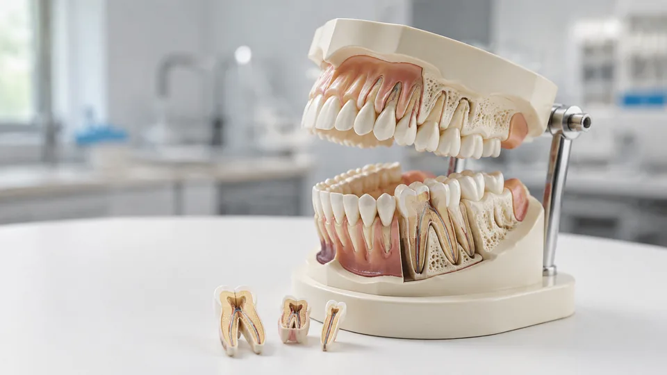
Dental anatomy becomes far easier to grasp once the jaw, roots, and surrounding bones stop living as flat shapes on a page. Rotate a transparent arch under a desk lamp and an impacted tooth suddenly makes spatial sense. Lift a mandible from a skull and the relationship between dentition and craniofacial structure feels much less abstract. That tactile clarity is why students, instructors, hygienists, and patient educators seek out dental anatomy models.
Models in this category serve surprisingly different purposes. Removable-tooth typodonts support identification and basic demonstrations, transparent pathology models reveal structures hidden below the gingiva, and full skulls provide broader context for the maxilla, mandible, foramina, and sutures. A common buying mistake is picking the most visually dramatic model without checking whether its scale, movable components, or displayed anatomy suit the lesson at hand.
Different buyers naturally have different priorities. A dental student may want individual teeth that can be removed and replaced, while a clinic might need large, obvious pathology features that patients can understand across a consultation desk. The Dentalmall Dental Demonstration Teeth Model earns the top overall position for its 28 removable teeth, separable jaws, and soft gingiva, but several specialized models below are better for pathology or craniofacial study.
Quick Answer
Best overall: The Dentalmall model offers the most useful balance of removable dentition, flexible gingiva, and clear viewing access. For explaining cavities, implants, root canals, and other conditions, the Ultrassist Transparent Dental Disease Teeth Model provides a more revealing view beneath the surface. Students studying wider cranial relationships should look toward the JENACAD Life-Size Human Skull Model, which pairs 55 numbered landmarks with a removable mandible and skullcap.
Short on time? The table gives a clean snapshot of all nine picks before the detailed reviews.
Best Dental Anatomy Models Comparison Table
| Product | Best For | Model Type | Key Feature |
|---|---|---|---|
| Dentalmall Demonstration Teeth Model | Overall dental study | Adult typodont | 28 removable teeth and soft gingiva |
| Ultrassist Dental Disease Model | Pathology education | Transparent jaw model | Displays roots, implants, decay, and bridges |
| JENACAD Life-Size Skull Model | Craniofacial study | Three-part skull | 55 numbered landmarks |
| Dental Caries Tooth Model | Large-group explanation | Enlarged pathology model | Highly visible common dental problems |
| Smile1000 Implant Teeth Model | Students and training | Implant typodont | 28 removable teeth |
| New Anatomy 15-Part Skull | Portable bone study | Mini exploded skull | 15 color-separated parts |
| Doctor Verified Life-Size Skull | Detailed skull-base review | Three-part skull | Numbered landmarks and magnetic joints |
| Transparent Dog Tooth Model | Canine veterinary dentistry | Transparent animal jaw | Visible canine tooth arrangement |
| Transparent Cat Tooth Model | Feline veterinary dentistry | Compact transparent jaw | Species-specific feline dentition |
The closer reviews below explain where each model shines and which limitations matter during real study or teaching sessions.
1. Best Overall: Dentalmall Dental Demonstration Teeth Model
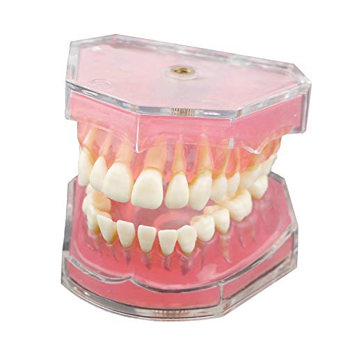
The Dentalmall model lands in the top spot because its components support active study rather than passive display. All 28 teeth are removable, so a student can lift one out, inspect its form, and return it to the arch. The translucent, soft rubber gingiva bends around the teeth instead of resembling a hard pink shelf, while the tooth material is intended to approximate the firmness of natural teeth.
Upper and lower jaws detach, giving a clearer route to lingual and occlusal surfaces. That matters during a late-night morphology review when twisting an attached model toward the light becomes a clumsy nuisance. A clear acrylic cover also helps expose the underlying arrangement visually. This is a teaching typodont rather than a pathology display, making it particularly useful for tooth identification, basic occlusion demonstrations, and doctor-patient conversations.
Pros
- Includes 28 individually removable teeth
- Soft, translucent gingiva allows easier tooth removal and observation
- Upper and lower jaws can be separated for closer viewing
- Clear outer structure reveals anatomy that opaque models can hide
- Balances student study, teaching, and patient demonstration uses
Cons
- Does not present the range of diseases shown by dedicated pathology models
- Small removable teeth require organized handling to prevent pieces from being misplaced
This is the strongest fit for students who want to handle individual teeth or instructors who need a flexible general-purpose demonstration piece. Clinics focused mainly on explaining implants, decay, and root treatment may get clearer mileage from the Ultrassist pathology model. For foundational dentition study, though, Dentalmall offers the most adaptable feature set among these nine picks.
To buy this product, click here.
2. Best for Pathology Education: Ultrassist Transparent Dental Disease Teeth Model

Spoken explanations can become a fog of unfamiliar terms, especially after someone hears “impacted tooth,” “cyst,” and “root canal” in the same appointment. Ultrassist addresses that communication gap with a transparent, natural-size adult dental model showing multiple pathological and restorative examples. The listing identifies cavities, pulp decay, a dental cyst, root canal treatment, bridges, implants, and an impacted tooth among its visible features.
The model opens as far as 290 degrees, creating a broad sightline across the upper and lower arches. Detachable components add an interactive layer, while clear acrylic exposes roots and implanted structures that conventional gum-colored models conceal. On a consultation desk, that visibility lets an educator point to the relevant feature instead of sketching a hurried cross-section on paper, which is precisely where this model earns its keep.
Pros
- Displays several common dental conditions and restorations in one model
- Transparent construction provides a direct view of roots and internal structures
- Opens widely for access to both dental arches
- Includes detachable components for closer explanation
- Natural-size format fits conveniently on a desk or clinic shelf
Cons
- Dense pathology features can feel busy during introductory tooth-identification lessons
- Natural-size details are less visible to students seated across a large classroom
Dental offices, hygiene instructors, and students reviewing pathology are the clearest audience. Its strength lies in showing several conditions within a compact model, rather than reproducing an ordinary healthy mouth. Buyers seeking repeated removal of every tooth should favor Dentalmall or Smile1000, while anyone explaining treatment relationships through clear acrylic will find Ultrassist unusually practical.
To buy this product, click here.
3. Best for Craniofacial Study: JENACAD Life-Size Human Skull Model

Teeth make more sense when studied within the architecture that supports them. This life-size JENACAD skull places the maxilla and movable mandible in broader cranial context, allowing dental and anatomy students to trace relationships among dentition, sutures, foramina, and adjacent bones. Fifty-five numbered landmarks create built-in prompts for self-quizzing, and red-painted sutures stand out crisply against the bone-colored PVC.
The three-part construction separates into a magnetic skullcap, skull base, and mandible. During practical review, a learner can remove the cap, turn the base upward, and follow a numbered landmark into a structure that looked puzzling in a flat atlas. The package also includes a printed study card and access to supplementary PDF and video materials. Its spring-supported jaw moves for examining tooth position, occlusion, and mandibular relationships, though the connectors call for gentle handling.
Pros
- Life-size proportions provide realistic craniofacial context
- Features 55 numbered landmarks for structured self-testing
- Removable skullcap and mandible expose internal and inferior anatomy
- Red sutures improve visual separation between cranial bones
- Includes printed and digital study resources
Cons
- Broader skull anatomy may be excessive for buyers focused solely on individual teeth
- Spring-supported jaw should be moved gently during frequent demonstrations
This is a smart pick for dental, medical, nursing, and anatomy students who need the whole craniofacial picture. Its roughly 7.5 by 5.5 by 6.5-inch body and listed weight of about two pounds make it substantial enough for tabletop instruction. A typodont remains better for removing individual teeth, but the JENACAD model offers far richer spatial context for the maxilla, mandible, and skull base.
To buy this product, click here.
4. Best for Visible Dental Problems: Dental Caries Tooth Anatomy Model

Tiny, natural-size pathology details can disappear from view the moment a model crosses a classroom. This dental caries model takes the opposite approach by enlarging premolars and molars, making common oral problems easier to see. Its displayed examples include cavities, abscesses, crowns, fillings, plaque, gingivitis, periodontitis, decay, bone loss, and related changes.
That bold presentation suits a hygiene lesson where several people need to identify the dark cavity or receding support without crowding around the instructor’s hands. It also gives clinicians a straightforward visual aid for explaining how apparently separate issues affect the tooth and surrounding tissue. The model is described as durable and easy to clean, store, and display, helpful traits for an item that may travel repeatedly between a cabinet and teaching table.
Pros
- Enlarged posterior teeth make pathology easier to see
- Shows a broad selection of common dental and periodontal problems
- Useful for classroom teaching and patient-facing explanations
- Offers a tactile alternative to flat posters and diagrams
- Compact display format is easy to store between lessons
Cons
- Focuses on selected premolars and molars rather than a complete adult dentition
- Exaggerated scale is less suitable for judging natural tooth-to-jaw proportions
Choose this model when visibility outranks completeness. It is particularly persuasive for health classes, hygiene instruction, and conversations about how plaque, decay, and bone loss appear in relation to dental structures. Students memorizing every tooth position need a full arch instead, but an educator tired of passing around a tiny transparent model may appreciate this model’s big, unmistakable details.
To buy this product, click here.
5. Best Value Typodont: Smile1000 Dental Implant Teeth Model

For learners who care more about removable dentition than elaborate color coding, the Smile1000 model offers a focused setup. It contains 28 removable teeth seated within translucent, soft rubber gingiva. Pulling a tooth free gives students a direct view of its position and shape, while the standard implant-model format supports basic demonstrations and doctor-patient communication.
This pick feels best suited to repeated desk study, where a student wants to point, remove, compare, and replace without unpacking a multi-part skull. The flexible gingiva should make those interactions more natural than rigid plastic gums. Its scope stays intentionally narrow, however. It is a training and teaching model rather than an enlarged pathology specimen or a numbered cranial reference, which keeps the learning experience refreshingly direct.
Pros
- Provides a complete set of 28 removable teeth
- Soft rubber gingiva supports hands-on tooth demonstrations
- Translucent material improves visibility around the dentition
- Suitable for student training and patient communication
- Focused design avoids unrelated cranial detail
Cons
- Provides fewer listed study aids than the top-ranked Dentalmall model
- Does not display a broad collection of pathological conditions
Smile1000 makes sense for students, dental assistants, and educators seeking a straightforward removable-tooth model without moving into a full skull system. It competes closely with the Dentalmall pick, although Dentalmall’s listed detachable jaws and acrylic cover give it a broader overall package. Select Smile1000 when the central task is handling and identifying teeth within a standard arch.
To buy this product, click here.
6. Best Compact Skull Model: New Anatomy 15-Part Color Exploded Skull Model
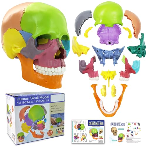
A palm-sized skull has obvious appeal for students whose study space is already buried under laptops, atlases, and half-finished coffee. This compact model separates into 15 colored parts representing paired and unpaired bones involved in the mouth, face, nasal cavity, and orbit. The listing identifies components such as the maxillae, cheekbones, nasal bones, palatine bones, vomer, mandible, and hyoid.
Color separation makes individual bones pop visually, while connectors allow the skull to be taken apart and reassembled along simplified sutures. That puzzle-like interaction can help a learner understand which structures form the bony scaffold around the oral cavity. It also depicts teeth, canals, sockets, eye openings, the internal nasal cavity, and the endocranial space. An illustrated guide accompanies the model, adding structure to a portable review session.
Pros
- Separates into 15 components for active bone study
- Color coding helps distinguish neighboring craniofacial bones
- Compact size is convenient for transport and small desks
- Includes an articulating mandible and complete visible dentition
- Supplied with an illustrated study guide
Cons
- Miniature scale makes fine landmarks harder to inspect than on a life-size skull
- Simplified suture connections provide less realistic articulation than natural cranial boundaries
This model suits visual learners who retain anatomy by assembling it piece by piece. Art students and anatomy learners can also use it to explore facial structure without dedicating a large shelf to a full-size skull. Dental students needing close tooth morphology should select a typodont, but this little skull is compelling for understanding how the mandible, maxilla, palate, and surrounding bones fit together.
To buy this product, click here.
7. Best for Skull-Base Landmarks: Doctor Verified 55-Landmark Life-Size Skull Model

Skull-base anatomy has a habit of turning tidy textbook diagrams into a maze once the specimen rotates. This full-size model places numbers on 55 landmarks, including structures around the skull base and foramina, so students can quiz themselves from multiple viewing angles. Downloadable PDF materials provide a reference for checking answers after that first uncertain pass.
Three detachable sections include the calvaria, skull base, and mandible. Strong embedded magnets secure the cap while preserving quick access to the interior, and the movable jaw supports observation of dentition and mandibular mechanics. High-density PVC construction is intended for long-term academic use. For a dental learner connecting the oral cavity with nerve pathways and neighboring cranial structures, those details carry far more teaching value than a decorative skull with a fixed jaw.
Pros
- Uses 55 numbered landmarks to guide structured review
- Includes detailed numbering around the skull base
- Full-size proportions support realistic spatial study
- Magnetic skullcap allows convenient internal access
- Movable mandible displays jaw and dental relationships
Cons
- Lacks the removable individual teeth found on dedicated typodonts
- Number markings may be visually distracting for art reference or clean patient displays
Students preparing for anatomy practicals will gain the most from this model, especially where skull-base relationships and mandibular anatomy share the syllabus. The JENACAD skull adds red sutures and a broader mix of learning media, while this option emphasizes secure magnetic construction and numbered structural review. Neither replaces a tooth-specific model, but both provide context a detached dental arch cannot show.
To buy this product, click here.
8. Best for Canine Veterinary Dentistry: Transparent Dog Tooth Model

Human typodonts are little help when the patient has prominent canines and a species-specific dental arrangement. This transparent blue dog model presents canine incisors, canines, premolars, and molars in a compact jaw measuring approximately 4.13 by 2.95 by 1.96 inches. The clear material lets veterinary learners look through the outer structure rather than relying entirely on exposed crowns.
Its brightest use case is client education in a veterinary clinic. Place it beside a dental chart and a pet owner can follow where a tooth sits as the clinician describes an oral health concern. The sleek blue construction also looks polished on a shelf, but its educational role carries more weight than the decorative appeal. Veterinary colleges, laboratories, and animal-health exhibitions are the natural settings for this specialized model.
Pros
- Represents dog dentition rather than adapting a human dental model
- Transparent construction supports internal visualization
- Shows major canine tooth groups in their jaw arrangement
- Compact footprint fits easily on clinic desks
- Useful for veterinary teaching and pet-owner communication
Cons
- Species-specific anatomy makes it unsuitable for human dental courses
- Small dimensions limit visibility during room-wide demonstrations
Veterinary students and practices should choose this over a human typodont whenever canine dentition is the actual subject. Its modest dimensions make one-to-one discussion easy, though instructors addressing a large lecture hall may still need projected images or an enlarged companion visual. For a veterinary consultation desk, the transparent blue jaw offers a quick and memorable point of reference.
To buy this product, click here.
9. Best for Feline Veterinary Dentistry: Transparent Cat Tooth Model

Feline dentition deserves its own reference, particularly because substituting a dog jaw muddies the size, spacing, and arrangement students are trying to learn. This blue transparent cat model displays incisors, canines, premolars, and molars within a species-specific jaw. At approximately 3.35 by 1.97 by 1.38 inches, it is the most compact specialized model in this roundup.
Picture a veterinary clinician trying to explain a hard-to-see tooth while an anxious cat waits in its carrier. A tidy model turns that awkward conversation into a visible map that can be rotated under the exam-room light. It also serves veterinary students studying feline oral anatomy and research teams needing a small demonstration reference. The transparent presentation is visually striking, though the petite scale rewards close viewing.
Pros
- Provides a dedicated representation of feline dentition
- Transparent blue material exposes internal tooth positioning
- Very compact size makes storage and transport simple
- Suitable for veterinary classrooms, clinics, and laboratories
- Supports clearer oral-anatomy discussions with cat owners
Cons
- Compact scale makes small structures difficult to see from a distance
- Offers limited usefulness outside feline veterinary education
This model belongs in a feline-focused classroom or veterinary practice rather than a human dental lab. Buyers comparing it with the dog version should select by species, since the cat model is appreciably smaller and represents different dentition. Used at arm’s length during consultation or personal study, its transparency and tiny footprint make it a handy specialist tool.
To buy this product, click here.
How to Choose a Dental Anatomy Model
Match the Model to the Lesson
Begin with the action the learner needs to perform. Tooth morphology and position call for a typodont with removable teeth. Pathology discussions benefit from transparent acrylic or enlarged diseased structures. Craniofacial courses need a skull with an articulated mandible, visible sutures, and access to the skull base. Buying a full skull for a simple brushing demonstration creates clutter, while bringing a tiny dental arch to an osteology lesson leaves crucial relationships out of sight.
Scale and Viewing Distance
Natural-size models preserve realistic proportions and fit nicely beside a textbook or consultation monitor. Their weakness appears when ten students gather around one table and the roots vanish behind someone’s shoulder. Enlarged models sacrifice true scale to make cavities, plaque, and periodontal changes obvious. For individual study, realistic dimensions usually win. For group teaching, bold enlargement often communicates faster.
Removable Teeth and Detachable Parts
Removable components invite active learning. A tooth can be lifted, oriented, named, and replaced, creating a physical sequence that a photograph cannot reproduce. Detachable jaws expose lingual surfaces, while removable skullcaps open access to internal structures. More pieces also mean more organization. A small compartment case or labeled tray can prevent an enthusiastic study session from ending with a missing molar under the couch.
Transparency, Numbering, and Color Coding
Transparent materials reveal roots, implants, impacted teeth, and pathology beneath the surface. Numbers turn a model into a self-testing tool, though they can make it look busy during clean visual demonstrations. Color coding is especially valuable on exploded skulls because paired bones become easier to separate at a glance. Select the visual system that supports the task rather than assuming more markings always mean better teaching.
Articulation and Material
A movable mandible helps demonstrate jaw position and occlusal relationships, but its connectors deserve gentle handling. Soft silicone or rubber gingiva feels more forgiving during tooth removal than rigid material. Acrylic provides crisp transparency, while PVC is common in durable skull models. No material choice solves every need, so focus on the components that will be manipulated most frequently.
Human Versus Veterinary Anatomy
Species accuracy matters. Dog and cat jaws should not be treated as scaled versions of one another, and neither belongs in a human dental course. Veterinary buyers should choose the model matching the animals they study or treat. Mixed-animal clinics may benefit from separate canine and feline models because a side-by-side comparison can make species differences immediately visible.
Best Dental Anatomy Models FAQ
What Type of Dental Model Is Best for Students?
A removable-tooth typodont is the most versatile starting point for dental students studying tooth position, arch arrangement, and basic morphology. Students covering head and neck anatomy may also need a life-size skull with a removable mandible and labeled landmarks.
Are Transparent Dental Models Better Than Opaque Models?
Transparent models are better for viewing roots, implants, cysts, impacted teeth, and internal relationships. Opaque or gum-colored models can present normal surface anatomy more cleanly. The stronger choice depends on whether the lesson concerns hidden structures or external form.
Do Removable-Tooth Models Work for Procedure Practice?
Some are marketed for training, but buyers should check whether a listing specifically supports the intended exercise. A basic demonstration typodont may help with identification and handling without having the construction required for drilling, preparation, or other procedural simulations.
Is a Full Skull Necessary for Dental Anatomy?
It is useful rather than universally necessary. A skull shows how the teeth relate to the maxilla, mandible, palate, cranial base, foramina, and facial bones. Learners focused narrowly on individual teeth may find a typodont more efficient and easier to carry.
Should a Classroom Model Be Enlarged?
Enlargement helps groups see cavities, gingival changes, restorations, and bone loss from several feet away. Natural-size models are more appropriate for learning realistic proportions or conducting one-to-one explanations. Many teaching settings benefit from having one of each.
How Should Removable Dental Models Be Stored?
Keep the model dry, shield it from prolonged direct sunlight, and follow the manufacturer’s cleaning guidance. Count removable teeth before storage and use a covered tray or small organizer for loose pieces. Handle flexible gingiva and jaw springs gently to reduce unnecessary strain.
Can Dental Anatomy Models Replace Formal Instruction?
No. They are educational references that support spatial understanding, demonstrations, and review. They do not replace qualified instruction, supervised clinical training, approved course materials, or professional evaluation.
Final Verdict
Among these nine models, the Dentalmall Dental Demonstration Teeth Model is the strongest overall choice. Its 28 removable teeth, flexible translucent gingiva, detachable jaws, and clear cover create a balanced platform for individual study and everyday teaching. It is broad enough to remain useful across several lessons without becoming crowded with pathology markings or wider cranial structures.
Specialized needs point toward different winners. The Ultrassist Transparent Dental Disease Teeth Model is the sharper choice for explaining implants, cavities, bridges, and hidden root structures. Students tackling craniofacial anatomy should consider the JENACAD Life-Size Human Skull Model for its numbered landmarks, removable components, and visible sutures. Before buying, picture where the model will sit, how far away learners will be, and whether they need to remove teeth, inspect pathology, or study the entire skull. That practical match matters far more than choosing the model with the longest feature list.
