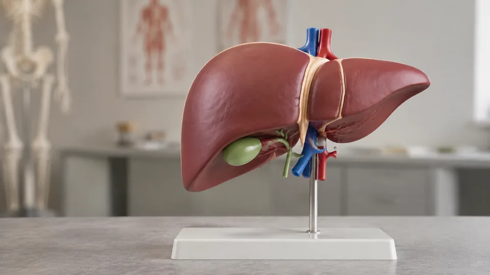
A liver model can turn a flat textbook diagram into something you can rotate, separate, point at, and finally understand. Yet these models differ sharply. Some reveal the liver’s relationship with the pancreas and duodenum, others magnify small landmarks, and pathology models show changes that a healthy-organ replica cannot. Buying the wrong type often leaves students staring at details they do not need or instructors missing the very structures they planned to teach.
Start with the learning goal. Removable components help with spatial anatomy, enlarged models improve visibility across a classroom, and silicone replicas provide a softer, more lifelike form for handling demonstrations. Buyers also need to check scale, represented structures, labeling, material, and stand design rather than choosing by color alone. For broad anatomy instruction, the faruijie Human Liver Anatomical Model is the strongest all-around choice here because its three-part design presents the liver alongside several closely related digestive structures.
Different settings call for different tools, so an anatomy student, hepatology clinic, nursing instructor, and science-minded gift buyer may land on four very different picks.
Quick Answer
The faruijie Human Liver Anatomical Model offers the best balance for general teaching, with three removable parts and visible pancreatic and hepatic vessels. Choose the Simedutech Pathology Liver Model for disease-focused explanations, or step up to the Simedutech Silicone Comparative Liver Model when side-by-side comparison of healthy and fatty livers is central to the lesson. The qzzXoq 1.5X Human Liver Anatomy Model is particularly useful when enlarged landmarks and numerical markers matter.
For a fast scan before the detailed reviews, the table below compares all nine selections by purpose, scale, construction, and defining feature.
Best Liver Anatomy Models Comparison Table
| Product | Best For | Scale And Material | Key Feature |
|---|---|---|---|
| faruijie Human Liver Anatomical Model | Overall anatomy teaching | Life-size PVC | Three-part hepatobiliary and digestive anatomy |
| Simedutech Pathology Liver Model | Pathology education | 1:1 PVC | Displays several hepatic diseases |
| qzzXoq 1.5X Human Liver Anatomy Model | Students studying landmarks | 1.5X PVC | Numerical markers and enlarged structures |
| Simedutech Silicone Comparative Liver Model | Comparative pathology | Three life-size silicone models | Healthy, alcoholic fatty, and non-alcoholic fatty livers |
| Hepatic and Biliary Vascular Model | Vascular and biliary study | Life-size model on base | Exposed vessels and bile ducts |
| Deluxe Human Liver Anatomical Model | Classroom display | 1.5X two-part PVC | Stable base and enlarged vascular anatomy |
| qzzXoq Liver Pancreas Duodenum Model | Guided independent study | Life-size three-part PVC | Numerical identification and instruction manual |
| Simedutech Healthy Liver Anatomy Model | Healthy-organ demonstrations | Life-size silicone | Flexible, latex-free construction |
| GIANT MICROBES Stuffed Liver Plush | Gifts and introductory education | 6-inch plush | Soft model with educational fact card |
The reviews below take a closer look at how each model fits real classrooms, study sessions, clinics, and educational displays.
1. faruijie Human Liver Anatomical Model: Best Overall

One reason this model rises to the top is its broader anatomical context. The three-part design helps an instructor show the relationships among the liver, duodenum, spleen, and pancreas rather than presenting the liver as an isolated red-brown shape. Its reproduced vessels include the hepatic portal vein, pancreatic vessels, and abdominal trunk, giving a student useful landmarks for tracing circulation and digestive anatomy during a tabletop review.
The PVC construction is intended to resist deformation and oxidation during long-term use, while computer-assisted color matching and hand-painted finishes distinguish neighboring structures. Picture a tutoring session where a student keeps confusing the pancreas with the nearby duodenum. Separating and reassembling these parts creates a much clearer spatial lesson than sliding a finger around a crowded page, which is precisely where this model earns its place.
Pros
- Three-part construction supports demonstrations of connected digestive structures
- Life-size format provides a practical anatomical reference
- Shows the hepatic portal vein, pancreatic vessels, and abdominal trunk
- Color differentiation makes neighboring structures easier to follow
- Durable PVC is suitable for repeated classroom or office handling
Cons
- Its multi-organ emphasis may be broader than buyers seeking liver-only surface anatomy need
- It does not provide the flexible feel of a silicone replica
This is the most balanced selection for anatomy classes, medical study groups, and patient-facing explanations that involve nearby digestive organs. Buyers concentrating on cirrhosis or fatty liver should choose a pathology model, while learners focused on tiny hepatic landmarks may prefer a magnified option. For general-purpose instruction, however, the removable pieces and visible vascular relationships offer unusually practical teaching value.
To buy this product, click here.
2. Simedutech Pathology Liver Model: Best for Disease Education

Pathology can feel painfully abstract when every condition is reduced to a paragraph and a tiny photograph. This 1:1 adult liver model gives disease-focused lessons a tangible center, displaying internal anatomy alongside cirrhotic septal nodules, bile duct obstruction, gallstones, and tumors. That combination suits instructors who need to move from normal structure into visible disease changes without swapping between several disconnected teaching aids.
A detachable stand adds welcome flexibility. Keep the model upright for a lecture or clinic display, then remove it when closer viewing angles are needed around a seminar table. Its lightweight PVC construction is easier to move than a heavy display specimen and is intended to tolerate frequent handling. During a patient explanation, a clinician can point to a bile duct obstruction directly, replacing a fog of unfamiliar terms with a visible route through the organ.
Pros
- Represents cirrhotic nodules, bile duct obstruction, gallstones, and tumors
- Uses a true-to-life 1:1 adult liver scale
- Detachable stand supports display and handheld demonstration
- Internal details add depth beyond a simple exterior replica
- Lightweight PVC suits repeated educational handling
Cons
- Pathological features make it less suitable as a standalone reference for a completely healthy liver
- It does not provide removable surrounding digestive organs
Nursing programs, health educators, clinics, and students covering hepatobiliary disease will get the most from this model. A foundational anatomy course may benefit from pairing pathology with a healthy replica so learners do not blur normal and abnormal structures. Compared with the larger comparative set below, this is a more compact route to covering several disease manifestations in one display.
To buy this product, click here.
3. qzzXoq 1.5X Human Liver Anatomy Model: Best for Students

If the goal is to identify liver landmarks without squinting, a 1.5-times enlarged model makes good sense. This qzzXoq design displays the lobes, ligaments, hilum, gallbladder, and bile duct system, with numerical markers supporting structured review. Those features suit the familiar study routine of pointing to a number, naming the structure aloud, and checking the reference before repeating the circuit.
The non-toxic PVC construction is easy to clean and compact enough to store between lessons, according to the listing. Distinct visual treatment helps separate major components at a glance, which can be valuable in a classroom where students gather around a demonstration desk. Rather than crowding six people around a life-size organ and hearing “Wait, which ligament?”, the enlarged surfaces give each learner a better viewing angle.
Pros
- 1.5X magnification improves visibility of anatomical landmarks
- Displays liver lobes, ligaments, hilum, gallbladder, and bile ducts
- Numerical markers support guided identification exercises
- PVC surface is described as easy to clean
- Useful for classroom, laboratory, and individual study settings
Cons
- Enlarged scale is less representative of the liver’s actual adult size
- Focuses on liver anatomy rather than relationships with the pancreas and duodenum
This model fits medical and nursing students who want clear, repeatable landmark practice, particularly before practical identification exercises. Educators can also use the magnification to make small structures more visible from nearby seats. Buyers who require life-size spatial reference should look elsewhere, but learners prioritizing clarity over exact scale will appreciate the larger format.
To buy this product, click here.
4. Simedutech Silicone Comparative Liver Model: Best Premium Set

Side-by-side comparison is where this premium set earns its keep. It includes three life-size models representing a normal liver, an alcoholic fatty liver, and a non-alcoholic fatty liver. An educator can place all three across a table and guide learners through visible differences without relying on memory between slides. That arrangement is particularly useful for nursing education, public-health presentations, and clinic conversations centered on liver health.
Each model uses soft, latex-free silicone that is flexible, skin-safe, and easy to clean, according to the product information. The texture and full-size form make the set more tactile than rigid PVC displays. Imagine rotating among stations in a clinical-skills workshop. Students can handle each representation, compare shape and texture, then return to the healthy model as a baseline. It is a focused, vivid lesson, though the three-piece set asks for more storage and budget than a single model.
Pros
- Includes normal, alcoholic fatty, and non-alcoholic fatty liver models
- Life-size pieces allow direct side-by-side comparison
- Flexible silicone provides a softer handling experience than rigid PVC
- Latex-free material suits repeated educational use
- Well matched to nursing programs, clinics, and health education events
Cons
- The three-model format requires more storage space than a single liver display
- Its specialized comparative focus may exceed the needs of introductory anatomy students
This set makes the strongest case for institutions that regularly teach fatty liver pathology and need a memorable visual comparison. It is harder to justify for a student seeking a basic study aid, especially since it does not center on removable ducts or surrounding organs. For recurring demonstrations where tactile comparison matters, the silicone construction and three distinct representations provide a clear advantage.
To buy this product, click here.
5. Hepatic and Biliary Vascular Model: Best for Vascular Anatomy

For buyers who prioritize the branching architecture inside the liver, this life-size model takes a more specialized path. It displays portal vein branches, hepatic veins, bile ducts, and cystic vessels, revealing relationships that can look like a tangled map in two dimensions. The exposed network supports lessons on hepatic circulation and biliary anatomy where a solid exterior model would hide the main attraction.
The model sits on a transparent Plexiglass base, allowing viewers to look through and around the vascular arrangement from multiple angles. That makes a difference during surgical education or an advanced anatomy session, where tracing one branch from origin to destination is the whole point. A learner can step around the base and follow a vessel’s course instead of mentally reconstructing depth from a flat illustration, bringing a notoriously dense topic into sharper focus.
Pros
- Clearly presents portal vein branches and hepatic veins
- Includes bile ducts and cystic vessels for biliary study
- Life-size format preserves realistic overall proportions
- Transparent base supports viewing from several angles
- Labeled structures aid medical teaching and demonstration
Cons
- Specialized vascular presentation is less useful for studying the liver’s solid external form
- The exposed branching design may be too advanced for basic introductory lessons
Medical students, anatomy instructors, and healthcare professionals studying hepatic circulation are the clearest audience. It also works as a striking professional display because its vessel network stays visible above the clear base. Beginners learning lobes and surface landmarks should start with a conventional organ model, while advanced learners trying to untangle portal and biliary pathways will find this option far more informative.
To buy this product, click here.
6. Deluxe Human Liver Anatomical Model: Best for Classroom Display

This two-part liver model combines 1.5X enlargement with a stable display base, a practical pairing for instructors who want an organ model ready at the front of the room. It shows the hepatic artery, portal vein, bile duct, and surrounding vascular network. The enlarged format keeps those details from disappearing when several students lean in around the same laboratory bench.
Its non-toxic PVC construction is sturdy and easy to clean, while the mounted presentation reduces the awkward juggling that happens when an instructor holds a loose organ in one hand and points with the other. Set it on a lectern or clinic desk, rotate attention among the colored structures, and continue the explanation without pieces rolling away. The two-part construction still adds interactive value, yet the stable base remains its strongest everyday convenience.
Pros
- 1.5X enlargement makes major structures easier to see
- Two-part design allows closer anatomical inspection
- Displays the hepatic artery, portal vein, and bile duct
- Stable base supports lectures and permanent display
- Durable PVC is suitable for regular classroom handling
Cons
- Mounted format is less portable than a compact loose model
- Magnification does not provide a true life-size spatial reference
Choose this model for a classroom, skills lab, or office where visibility and a tidy presentation matter more than portability. Students carrying a model between home and campus may prefer a smaller three-part option. Compared with the numbered qzzXoq model, this selection places greater emphasis on its two-part display and base, making it an appealing choice for instructor-led demonstrations.
To buy this product, click here.
7. qzzXoq Liver Pancreas Duodenum Model: Best for Guided Study

Independent study becomes much smoother when a model supplies a clear identification system. This life-size qzzXoq model combines numerical labels with an instruction manual, giving learners a route through its liver, pancreas, duodenum, spleen, and vascular details. It separates into three parts and reveals the external pancreas, pancreatic duct, celiac artery, and a major vein, making it useful for studying anatomical relationships rather than memorizing isolated names.
The hand-painted PVC uses distinct colors and can be cleaned after frequent handling. In a small study group, one person can remove a component while another follows the corresponding number in the manual, turning a passive review session into a brisk identification drill. That self-guided structure separates it from models that look impressive on a shelf but provide little help once the textbook closes.
Pros
- Three detachable parts support spatial anatomy lessons
- Numerical labels pair with an included instruction manual
- Shows the pancreas, pancreatic duct, duodenum, and spleen
- Represents celiac and major venous anatomy
- Hand-painted PVC is described as easy to clean
Cons
- The many represented structures can feel busy for learners seeking liver-only anatomy
- Rigid PVC offers less tactile realism than flexible silicone
This is a strong study-desk option for medical students, anatomy enthusiasts, and training programs that value numbered guidance. It overlaps with the faruijie model in broad anatomy and three-part construction, but its digital identification and manual make it particularly appealing for self-directed review. Buyers seeking pathology features or enlarged liver landmarks should select a more specialized model.
To buy this product, click here.
8. Simedutech Healthy Liver Anatomy Model: Best Flexible Replica

A rigid plastic model excels at crisp landmarks, but sometimes an educator wants a full-size organ representation that feels softer in the hands. This Simedutech model uses flexible, latex-free silicone and depicts a healthy adult liver’s normal shape, color, and structure. Its straightforward presentation establishes a clean baseline before an instructor introduces fatty liver, cirrhosis, tumors, or biliary disease.
The washable silicone construction suits medical training, nursing education, and patient demonstrations where the model may pass from hand to hand. Picture a clinical-skills workshop in which students assess the organ’s size and orientation before returning it to its anatomical position. The life-size format helps build that spatial awareness without the distraction of multiple detachable components, keeping attention on the healthy liver itself.
Pros
- Life-size form supports realistic spatial understanding
- Flexible silicone provides a tactile alternative to rigid PVC
- Latex-free construction broadens suitability for shared educational settings
- Healthy anatomy creates a clear baseline for foundational teaching
- Easy-clean material supports repeated handling
Cons
- Does not display the range of disease changes found on pathology models
- Lacks detachable surrounding organs and exposed vascular networks
This replica suits nursing labs, patient education, and introductory instruction where healthy anatomy and handling realism take priority. It is less useful for memorizing numbered landmarks or tracing individual vessels, so examination-focused students may prefer an enlarged labeled model. As a tactile reference for normal liver form, however, it fills a role that hard PVC displays cannot quite match.
To buy this product, click here.
9. GIANT MICROBES Stuffed Liver Plush: Best Educational Gift

Serious anatomy study has its place, but a little levity can open the door to curiosity. This soft liver measures approximately 6 by 4 by 2 inches and uses embroidered anatomical details rather than molded PVC structures. An included information card adds educational context, making the plush more meaningful than an ordinary novelty toy for young science fans, medical students, or healthcare professionals.
The compact size slips easily onto a desk, classroom shelf, or recovery gift basket, and its friendly expression can make a sensitive health conversation feel less intimidating. A child who recoils from a realistic organ replica may happily pick up this plush and ask what the liver does. That moment matters. Still, its cuddly shape serves broad engagement rather than detailed identification of ducts, vessels, and ligaments.
Pros
- Soft plush format makes liver education approachable
- Embroidered details provide a simple anatomical reference
- Includes an educational information card
- Compact size works well for desks, gifts, and informal demonstrations
- Suitable for science enthusiasts and healthcare-themed occasions
Cons
- Cannot support detailed study of internal liver structures
- Small plush proportions are unsuitable for life-size anatomical reference
This pick belongs in gift bags, introductory science activities, and relaxed educational settings rather than an advanced anatomy laboratory. It can spark a conversation or offer a comforting bit of humor, which is a different job from preparing someone for a practical examination. Choose it for accessibility and charm, then select one of the rigid or silicone models above when precise structure becomes the lesson.
To buy this product, click here.
How to Choose a Liver Anatomy Model
Match the Model to the Lesson
Begin with what the model must explain. A medical student reviewing the hepatic portal system needs visible vessels, while a nursing instructor discussing fatty liver benefits from healthy and pathological examples placed side by side. For a basic digestive anatomy lesson, a three-part model that includes the pancreas and duodenum provides valuable context. A specialized vascular model can be brilliant for advanced study yet confusing during someone’s first encounter with hepatic anatomy.
Think through a typical session before buying. If the model will sit at the front of a classroom while an instructor points from several feet away, enlargement and bold color separation are useful. If two students will quiz each other at a kitchen table before an exam, removable parts, numbers, and a reference guide may matter more than a handsome stand.
Scale and Size
Life-size models communicate the approximate adult organ scale and make it easier to understand how the liver occupies space in the upper abdomen. Enlarged 1.5X models trade that realism for clearer visibility. They can expose the hilum, ligaments, ducts, and vascular landmarks without making learners lean so close that everyone bumps heads around the desk.
Compact models are easier to carry and store, but small details may become harder to distinguish. Measure the intended shelf or cabinet and check the listing before ordering, especially for enlarged or multi-model sets. Scale should serve the lesson rather than becoming a decorative talking point.
Removable Parts and Surrounding Anatomy
Detachable parts help learners understand depth and anatomical relationships. A liver presented with the pancreas, spleen, and duodenum can clarify how structures sit near one another, while a two-part liver may reveal internal features without adding the visual clutter of a larger digestive system. Frequent removal also creates more handling at connection points, so classroom rules and careful storage remain sensible.
A fixed model can still be the better choice for lectures and display. Mounted pieces stay organized, take seconds to set up, and reduce the chance that a small component goes wandering after lab. Anyone who has searched a classroom floor for a missing plastic duct knows why that convenience counts.
Labels, Numbers, and Color Coding
Numbered structures work well for self-testing, provided the product includes a usable key or manual. Direct labels can accelerate demonstrations, while unlabeled models encourage recall when paired with a separate anatomy atlas. Bright color coding improves visual separation, but color should never be mistaken for a perfectly literal representation of living tissue.
Look for the landmarks tied to your curriculum. Those may include lobes, ligaments, the hilum, hepatic artery, portal vein, hepatic veins, gallbladder, and bile ducts. A long feature list has little value if it skips the structures on your study sheet.
PVC Versus Silicone
PVC models are common because they hold crisp molded detail, clean easily, and remain practical for repeated classroom use. Their firm surfaces suit numbered landmarks, detachable sections, and stable mounting. They feel plainly manufactured, though that rigidity often makes small features easier to point out.
Silicone provides flexibility and a softer tactile experience, which can help in handling demonstrations and clinical-skills education. It often costs more and may offer fewer sharply molded detachable structures. Choose silicone for touch and life-size form, or PVC for crisp visual instruction and component-based study.
Healthy Anatomy Versus Pathology
A healthy liver model gives beginners a dependable baseline. Pathology models add cirrhotic nodules, obstruction, tumors, gallstones, or fatty changes, depending on the specific design. Those features can make disease education much more concrete, but they should not blur a learner’s understanding of ordinary anatomy.
For recurring pathology lessons, separate healthy and diseased models allow the cleanest comparison. A single combined pathology display saves space and can cover several conditions efficiently. Neither format replaces formal medical instruction or clinical material, but each can make anatomical explanations easier to visualize.
Best Liver Anatomy Models FAQ
Is a Life-Size or Enlarged Liver Model Better?
A life-size model is better for learning realistic organ scale and spatial placement. An enlarged model is easier to see during group demonstrations and can make small ducts, vessels, ligaments, and hilum structures clearer. Students often benefit from enlargement, while clinical demonstrations may favor life-size proportions.
Which Structures Should a Good Liver Model Show?
That depends on the course, but common targets include the liver lobes, gallbladder, bile ducts, hepatic artery, portal vein, hepatic veins, ligaments, and hilum. Models used for digestive anatomy may also include the pancreas, duodenum, spleen, and nearby vessels.
Are Removable Liver Models Better for Studying?
Removable models are excellent for exploring spatial relationships and hidden structures. They also make active recall more engaging because learners can disassemble and rebuild the anatomy. Fixed models remain preferable for quick lectures, permanent displays, and environments where loose pieces could be misplaced.
Can a Pathology Liver Model Teach Normal Anatomy?
It can show basic liver form and selected internal structures, but represented diseases may distract beginners from the healthy baseline. For foundational teaching, start with a healthy model or use healthy and pathological examples together so the differences remain clear.
Is PVC or Silicone Easier to Maintain?
Both materials can be easy to clean when the manufacturer permits routine surface cleaning. PVC is rigid and holds painted detail well, while silicone is flexible and offers a softer feel. Follow the seller’s care directions and avoid harsh cleaning products that could damage paint or material.
Do Anatomy Models Use Realistic Colors?
Manufacturers frequently use distinct, educational colors to separate vessels, ducts, and neighboring organs. Those colors improve readability but may be more vivid or simplified than actual tissue. Treat color coding as a navigation aid and cross-reference formal anatomical resources.
Are Liver Anatomy Models Suitable for Patient Education?
Yes, many models can help healthcare professionals explain basic anatomy or visually identify a represented structure. The model should support a clear conversation rather than serve as a diagnostic or treatment tool. Healthy, pathology, and vascular models each suit different explanations.
What Is the Best Liver Model for a Medical Student?
A numbered, enlarged model is useful for landmark identification, while a detachable multi-organ model helps with spatial anatomy. The best choice follows the student’s syllabus. Check the required structures first, then decide whether labeling, removable parts, realistic scale, or pathology carries the most weight.
Final Verdict
Among these nine models, the faruijie Human Liver Anatomical Model is the strongest overall recommendation. Its life-size presentation, three removable parts, visible vessels, and inclusion of surrounding digestive structures make it useful across individual study, classroom teaching, and patient-facing explanation. It provides enough detail to move beyond a basic liver shape without narrowing the lesson to one disease or vascular pathway.
More specialized buyers have compelling alternatives. The Simedutech Pathology Liver Model places several hepatic conditions into one convenient display, while the premium Simedutech Silicone Comparative Liver Model supports vivid side-by-side fatty liver education. Students who need larger, numbered landmarks should favor the qzzXoq 1.5X Human Liver Anatomy Model. Before ordering, compare the structures on the listing with your curriculum, confirm available storage space, and decide whether scale, detachable anatomy, pathology, or tactile realism will carry the lesson forward.
