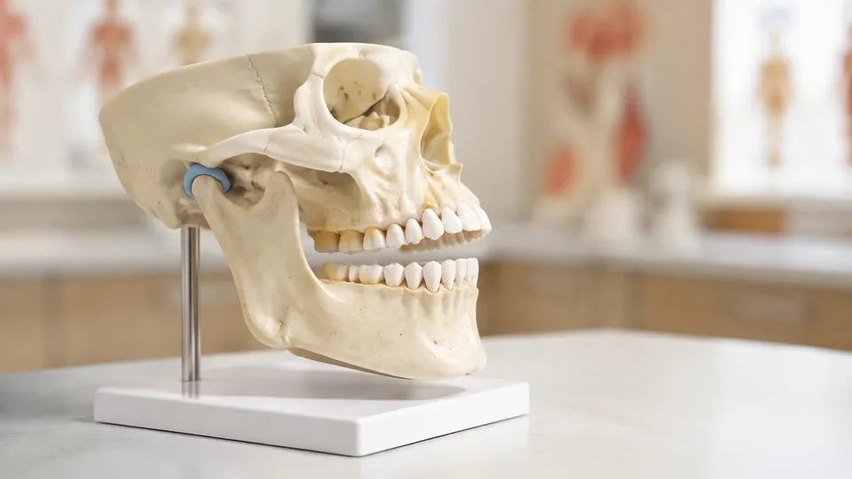
The jaw looks straightforward in a textbook until you try to trace how the mandible meets the temporal bone, how the teeth align, or where chewing muscles pull during movement. Suddenly, a tidy illustration becomes a maze of shallow fossae, narrow processes, hidden tooth roots, and structures tucked behind the ramus. A physical model lets students rotate that puzzle under a desk lamp, open the jaw, and study relationships that flat images tend to flatten further.
Jaw anatomy models vary far more than their product photos suggest. Some are full-size skulls that place the mandible in its cranial context, while others concentrate on teeth, removable components, or visible muscle anatomy. Common buying mistakes include selecting a tiny desktop model for detailed coursework, assuming every movable jaw is removable, or buying an unlabeled replica when guided identification would be more useful. Scale, articulation, removable parts, visible landmarks, and the intended learning environment matter most.
Different learners naturally have different priorities. A dental student may care about dentition and muscles of mastication, an anatomy instructor may need a life-size model visible across a lab table, and an artist may prefer clean bone surfaces without printed numbers. The Axis Scientific skull with masticatory muscles is the strongest overall option here because it combines realistic cranial structure with four major chewing muscles and an articulating mandible.
Quick Answer
Best overall: The Axis Scientific Life-Size Human Skull with Masticatory Muscles offers the most useful jaw-focused teaching package, especially for mastication and dental anatomy. For numbered study, the Doctor Verified 55 Number Labeled Skull provides direct landmark references. Buyers seeking a straightforward life-size model without printed prompts should look at the Human Skull Model for Anatomy, while the Alikeke miniature is the easiest option to fit into a cramped study corner.
For a fast scan before the full reviews, the table below compares the nine selected models and their most useful features.
Best Jaw Anatomy Models Comparison Table
| Product | Best For | Scale or Size | Jaw-Focused Feature |
|---|---|---|---|
| Axis Scientific Skull With Masticatory Muscles | Overall jaw mechanics | Life-size | Four chewing muscles and articulating jaw |
| Doctor Verified 55 Number Labeled Skull | Guided landmark study | Life-size, 1:1 | Numbered landmarks and movable mandible |
| Human Skull Model for Anatomy | Active recall | 7.5 x 5.5 x 6.5 inches | Removable, movable mandible without printed prompts |
| Axis Scientific Human Skull Model | General anatomy study | Life-size | Articulating mandible and labeled structures |
| Anatomy Lab Human Skull Model | Value-focused students | Life-size | Articulating mandible and removable calvaria |
| Dentalmall Dental Demonstration Teeth Model | Removable tooth study | Standard dental model | 28 removable teeth and separable jaws |
| Smile1000 Dental Implant Teeth Model | Dental communication | Standard dental model | Removable teeth and translucent gingiva |
| Alikeke Human Skull Anatomical Model | Compact study spaces | 4.5 x 3.5 x 3.5 inches | Articulated mandible and full set of teeth |
| GPI Anatomicals Clear Canine Jaw Model | Veterinary dental anatomy | 4.25 x 3 x 2 inches | Transparent jaw showing canine tooth roots |
The detailed reviews below show where each model earns its place and which compromises matter during actual study or instruction.
1. Axis Scientific Life-Size Human Skull With Masticatory Muscles: Best Overall

Muscle anatomy gives this life-size Axis Scientific model a decisive advantage for studying jaw function. The temporalis, masseter, medial pterygoid, and lateral pterygoid are reproduced in their anatomical positions, connecting names on a lecture slide to visible structures around the mandible. For a student puzzling over which muscle elevates, protrudes, or shifts the jaw, that spatial context can make the mechanics feel much less abstract.
The skull is cast from an actual human specimen, according to the listing, so its sutures, contours, and bony landmarks preserve realistic surface detail. Its mandible articulates to demonstrate opening and closing alongside the chewing muscles. A full-color manual uses photographs of the model itself, which is handy when a study session stalls over a structure that looks different from the simplified drawing in a course atlas.
Pros
- Shows the temporalis, masseter, medial pterygoid, and lateral pterygoid muscles
- Life-size skull provides valuable cranial context for the mandible
- Articulating jaw helps demonstrate basic movement and muscle relationships
- Cast from an actual human skull for realistic bony contours
- Includes a full-color manual with photographs of the model
Cons
- More specialized than a plain skull for students focused strictly on bone identification
- Hand-painted muscle structures require more careful handling than a basic one-piece teaching aid
This is the clearest choice for dental students, anatomy instructors, speech-language pathology learners, and anyone concentrating on mastication. Picture an instructor explaining the pterygoids to a small group: instead of pointing vaguely behind the jaw on a poster, the model puts those muscles where learners can inspect their orientation. Artists seeking bare bone contours may prefer the unnumbered model, while basic osteology students may find a simpler skull sufficient. For jaw mechanics, however, this model supplies the most meaningful visual information among the nine picks.
To buy this product, click here.
2. Doctor Verified 55 Number Labeled Life-Size Skull Anatomy Model: Best for Guided Landmark Study

Numbered landmarks can save a learner from constantly shifting attention between a skull, an atlas, and a page of handwritten notes. This life-size model marks 55 features, including structures around the skull base, and provides downloadable study material for matching each number to its anatomical name. That guided format suits early coursework, tutoring sessions, and practical-exam preparation where rapid identification is the main goal.
The model separates into the calvaria, skull base, and mandible. Strong embedded magnets hold the cranial components together, while the movable lower jaw displays dental alignment and basic articulation. High-density PVC gives it the wipe-clean practicality expected from a classroom model. Imagine passing it down a lab bench as each student locates a landmark. The numbered surface keeps everyone oriented rather than leaving half the group staring at the wrong opening.
Pros
- Features 55 numbered anatomical landmarks
- Life-size, 1:1 format supports realistic spatial study
- Movable mandible demonstrates jaw position and dentition
- Three-part construction provides access to the cranial cavity and skull base
- Includes access to downloadable study references
Cons
- Printed numbers can become visual prompts during unassisted recall practice
- Does not display the muscles responsible for mandibular movement
Students who learn well through numbered identification will get more immediate direction here than from a blank skull. It also makes sense for instructors who need to call out a structure and have several learners find the same spot quickly. Those preparing for an unnumbered lab practical should eventually cover the numbers or switch to a clean model for retrieval practice. Compared with the muscle-equipped Axis Scientific pick, this one favors broad landmark recognition over functional anatomy.
To buy this product, click here.
3. Human Skull Model for Anatomy, Life-Size 3-Part Skull: Best for Active Recall

Blank bone surfaces make this skull especially useful once the flash cards are memorized and it is time to identify structures without clues. There are no printed numbers or color-coded regions, so learners must recognize the mandible, facial bones, teeth, orbital features, and cranial contours from shape and position. That clean appearance also suits portrait artists who want a reference without bright markings cutting across the bone.
Measuring 7.5 by 5.5 by 6.5 inches, the PVC model approximates adult skull scale while remaining manageable on a desk. Its cranial cap attaches with magnets and pins, and the spring-connected mandible can be opened, repositioned, or removed. A paper manual and online learning access provide guidance away from the model surface. During a late-night study drill, a partner can point to random landmarks while the clean replica offers no easy hints.
Pros
- Unnumbered surface supports genuine active-recall practice
- Removable, movable mandible permits closer jaw inspection
- Life-size proportions provide useful spatial context
- Magnetic three-part construction allows access to internal cranial anatomy
- Includes paper and online study resources
Cons
- Beginners may need to consult the manual frequently without labels on the model
- Spring-connected articulation does not reproduce the complete biomechanics of the temporomandibular joint
This skull fits independent students, practical-exam preparation, classroom quizzes, and observational drawing. It is particularly appealing as a second-stage learning tool after a student has used numbered diagrams to become familiar with basic terminology. Complete beginners who want every landmark identified directly on the surface may prefer the Doctor Verified model. Buyers concentrating on chewing muscles should move toward the overall pick, but clean osteology practice is where this unnumbered model shines.
To buy this product, click here.
4. Axis Scientific Human Skull Model for Anatomy: Best for General Anatomy Study

For learners who need one skull to cover the jaw, cranial cavity, sutures, and skull-base anatomy, this Axis Scientific model strikes a useful middle ground. The three-part format consists of an articulating mandible, skull base, and removable calvarium. Opening the model reveals features that disappear from view on fixed replicas, giving osteology study more depth than a decorative skull can offer.
External and internal structures are identified, and the included full-color manual uses actual model photographs rather than unrelated generic illustrations. That detail matters during self-study because a landmark’s shape and viewing angle match the object sitting in front of you. The life-size format also works well in a small teaching group, where a student can turn the skull sideways and trace the relationship between the mandibular ramus and surrounding cranial structures.
Pros
- Life-size design supports realistic scale and spatial relationships
- Articulating mandible provides a practical view of jaw placement
- Removable calvarium exposes internal cranial structures
- Includes identified external and internal anatomical features
- Photo-based manual corresponds directly to the supplied model
Cons
- Lacks reproduced chewing muscles for detailed functional demonstrations
- Broader skull coverage may be more than buyers need for tooth-focused instruction
This option is well suited to anatomy and physiology students, educators, forensic anthropology learners, and patient-education settings that need a broad skull reference. Its advantage over dental typodonts is context: the teeth and mandible sit within the complete facial skeleton rather than floating as isolated arches. Learners interested primarily in removable teeth should choose a dental model, while those studying muscle action will gain more from the Axis Scientific muscle skull.
To buy this product, click here.
5. Anatomy Lab Human Skull Model: Best Value Full-Size Model

A straightforward full-size skull remains one of the most versatile tools for introductory anatomy, and the Anatomy Lab model keeps its feature set focused. It includes a removable calvaria, skull base, and articulating mandible, giving students access to the primary regions expected from a basic three-part model. There are no specialized muscle reproductions or removable individual teeth to complicate routine bone study.
The model is positioned for classrooms, clinical teaching spaces, artists, and independent learners who want adult-scale proportions. Anatomy Lab states that its models are reviewed for anatomical accuracy by its US-based medical team. Set on a dorm-room desk, it offers enough size to study the jaw and facial skeleton clearly, yet it can still be disassembled when a closer look at the internal base is needed.
Pros
- Life-size proportions suit introductory anatomical orientation
- Articulating mandible supports basic jaw demonstrations
- Removable calvaria expands study beyond external skull surfaces
- Simple three-part construction is approachable for beginners
- Useful across student study, classroom teaching, and art reference
Cons
- Provides fewer specialized jaw features than muscle or dental models
- Individual teeth are not described as removable
This model makes sense for buyers who want a traditional skull replica without paying for features outside their syllabus. It handles basic mandible positioning, facial-bone relationships, and skull orientation well. A dental learner practicing tooth placement will find the Dentalmall or Smile1000 designs more relevant, and advanced jaw-function lessons call for visible masticatory muscles. For broad introductory study, the Anatomy Lab skull remains a sensible, uncluttered option.
To buy this product, click here.
6. Dentalmall Dental Demonstration Teeth Model: Best for Removable Tooth Study
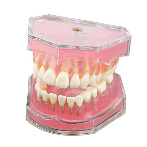
Jaw study sometimes means looking beyond the mandible as a bone and concentrating on what sits inside the dental arches. The Dentalmall model has 28 removable teeth, translucent soft rubber gingiva, and upper and lower jaws that detach. Pulling an individual tooth changes the lesson from looking at a row of white crowns to inspecting placement and orientation one component at a time.
The model combines simulated teeth with flexible silica-gel material and a clear acrylic cover. Its tooth-centered format makes it more relevant to dental demonstrations and study than to cranial osteology. Picture an instructor explaining tooth positions at chairside or a student organizing the removable teeth before returning them to the arches. That interaction delivers a different kind of understanding from opening and closing a skull’s spring-held jaw.
Pros
- Includes 28 individually removable teeth
- Upper and lower jaws can be separated for closer inspection
- Translucent gingiva leaves underlying placement easier to observe
- Flexible silica-gel construction provides a softer gum representation
- Clear acrylic cover helps protect the assembled model during storage
Cons
- Does not show the jaw in relation to the complete skull
- Removable components create more small pieces to organize and store
Dental students, educators, and buyers concentrating on dentition will find this model more practical than a fixed-tooth skull. It is especially useful where removing and replacing teeth forms part of the demonstration. Learners studying the mandibular condyle, coronoid process, skull base, or chewing muscles should select a full cranial model instead. This is a focused typodont-style teaching aid, and that narrow focus is precisely why it earns a spot.
To buy this product, click here.
7. Smile1000 Dental Implant Teeth Model With Removable Teeth: Best for Dental Communication

Clear demonstrations often work better than a flurry of technical terms, particularly when someone is trying to understand tooth position or general implant concepts. The Smile1000 model is presented for teaching, training, and doctor-patient communication. It includes 28 removable teeth and translucent, soft rubber gingiva, allowing an educator to point to a physical structure rather than asking a viewer to mentally decode a flat diagram.
Its compact dental format keeps attention on the arches and teeth instead of the broader cranium. During a tutoring session, a learner can remove a tooth, inspect its orientation, and return it to the model while discussing neighboring structures. The design has fewer documented details than the full-size skull picks, but its simplicity can be welcome where the lesson needs to stay centered on dental arrangement.
Pros
- Includes 28 removable teeth for interactive study
- Translucent gingiva helps reveal underlying tooth placement
- Soft rubber gum material supports clear dental demonstrations
- Suitable for teaching, training, and general patient communication
- Focused dental format is easier to handle than a full skull
Cons
- Offers limited reference for mandibular and cranial bony landmarks
- The listing provides fewer structural specifications than several models in this roundup
Choose this model for straightforward tooth demonstrations, dental tutoring, or visual communication where a complete skull would distract from the immediate topic. It is less appropriate for an anatomy practical covering the ramus, condyle, or muscle attachments. Compared with the Dentalmall model, both options emphasize removable teeth and soft gingiva, while the Smile1000 listing places particular emphasis on teaching and communication. Buyers should match that narrower purpose to the lesson they actually plan to deliver.
To buy this product, click here.
8. Alikeke Human Skull Anatomical Model: Best Compact Option

Limited shelf space can turn a life-size skull into an awkward roommate. At 4.5 by 3.5 by 3.5 inches, the Alikeke model condenses basic skull and jaw anatomy into a form that fits beside a laptop or slips into a teaching bag. It still includes a full set of teeth, removable skull cap, and articulated mandible, which gives the miniature more interactive value than a solid decorative replica.
The model uses PVC and is aimed at introductory education, display, and art reference. Its small scale naturally reduces how clearly tiny landmarks can be represented, so expectations should stay realistic. For a quick overview before class, though, being able to turn the jaw in your hand can beat pinching and rotating a digital image on a phone screen.
Pros
- Compact dimensions fit small desks and crowded shelves
- Articulated mandible provides basic movement
- Removable skull cap adds access to the upper cranial area
- Includes a complete visible set of teeth
- PVC construction is practical for routine handling
Cons
- Miniature scale limits visibility of fine anatomical landmarks
- Less suitable than a life-size skull for group demonstrations
This is the pick for portability, casual reference, compact displays, and students with very little room. It can introduce the broad relationship between the mandible, dentition, and cranium without occupying half a bookcase. Serious dental or medical study generally benefits from a life-size model with clearer contours, while instructors addressing a room will need something larger. As a personal desktop reference, the Alikeke offers useful movement in a remarkably small footprint.
To buy this product, click here.
9. GPI Anatomicals Clear Canine Jaw Model With Teeth: Best for Veterinary Anatomy

Human anatomy models dominate this category, but veterinary students and clinic educators need a very different jaw. The GPI Anatomicals canine model uses clear construction to expose the roots of incisors, canine teeth, premolars, and molars. Its hinged jaw opens and closes, and the sections can be separated for a closer view, turning structures normally buried inside bone into visible teaching points.
The jaw measures 4.25 by 3 by 2 inches and comes with an informational card. That compact format suits a veterinary office counter or a small classroom table, where an educator can show a pet owner why the visible crown is merely part of the tooth. The transparent body is the star here. Tooth roots remain visible from several angles without taking the model apart piece by piece.
Pros
- Transparent construction reveals canine tooth roots
- Displays incisors, canine teeth, premolars, and molars
- Hinged jaw opens and closes for basic demonstration
- Jaw separates for closer examination
- Includes an informational reference card
Cons
- Canine anatomy is irrelevant for buyers seeking a human jaw model
- Compact dimensions reduce visibility during large-group teaching
Veterinary students, animal-science educators, and veterinary clinics are the clear audience for this specialized model. In a consultation room, the exposed roots can communicate far more quickly than pointing at a tiny radiographic image. Human anatomy students should skip it because canine dentition and jaw proportions answer a different set of questions. Within its intended veterinary niche, however, the transparent design offers a view none of the human skull models can provide.
To buy this product, click here.
How to Choose the Best Jaw Anatomy Model
Start With the Anatomy You Need to Learn
“Jaw anatomy” can mean several different lessons. Osteology students usually need the mandibular body, ramus, angle, coronoid process, condylar process, foramina, and the jaw’s relationship to the skull. Dental learners may care more about tooth arrangement, roots, and gingiva, while functional anatomy courses place greater emphasis on the masseter, temporalis, and pterygoid muscles. A full skull gives broad context, a typodont concentrates attention on teeth, and a muscle model helps explain motion.
Think about the actual moment of use. A student preparing for a bone-identification practical gains little from removable teeth if the exam asks about cranial landmarks. A dental educator explaining tooth placement may find a heavy full skull unnecessarily cumbersome. Matching the model to the syllabus prevents an impressive-looking feature from becoming expensive clutter.
Life-Size Versus Miniature Models
Life-size skulls are usually easier for studying small contours and showing structures to two or three people gathered around a table. They preserve realistic spatial relationships and feel more natural when comparing the mandible with the face and cranial base. Their downside is obvious the moment textbooks, a laptop, and coffee already cover the desk: adult-scale models demand real storage room.
Miniature models trade fine detail for portability. They work well as quick visual references, compact art aids, and introductory teaching props. A small jaw may open and close perfectly well, yet tiny foramina and shallow depressions will still be harder to distinguish. For demanding coursework, prioritize readable anatomy over convenience unless the model will travel frequently.
Articulating Versus Removable Mandibles
An articulating jaw demonstrates opening and closing, but articulation alone does not guarantee that the mandible detaches. Removability is useful for turning the bone over, viewing its medial surface, and inspecting areas obscured while it is attached to the cranium. Students often discover this distinction after trying to see behind the ramus and finding that a spring or hinge blocks the angle they need.
These models remain simplified teaching tools. A movable mandible can illustrate position and general direction, but it does not fully reproduce living joint tissues, muscle activity, or complex temporomandibular motion. For functional lessons, visible chewing muscles provide more educational context than a bare hinged jaw.
Labels, Numbers, and Clean Surfaces
Numbered models are friendly guides during early learning. They let an instructor say “find number 32” and bring a group to the same landmark quickly. Their weakness appears during recall practice, where the printed number becomes a cue. Students may recognize the label while struggling to identify the underlying structure on an unmarked specimen.
Clean models reverse that trade-off. They are excellent for quizzes, active recall, photography, and drawing, but beginners must keep a manual or atlas nearby. One practical approach is to learn on a numbered model or reference sheet, then practice on an unnumbered skull once the names begin to stick.
Teeth, Gingiva, and Removable Components
Fixed teeth are adequate for general skull orientation and occlusal relationships. Removable teeth become useful for demonstrations centered on tooth position, arrangement, or individual components. Translucent gingiva can make underlying placement easier to see, while clear jaw construction can expose roots that opaque bone would hide.
More removable parts also mean more pieces rolling under desks. In a busy classroom, a model with 28 separate teeth needs a consistent storage routine. Buyers who rarely need to remove teeth may prefer a sturdy fixed dentition and a removable mandible instead.
Muscle Detail and Functional Anatomy
Visible muscle reproductions add major value for learning mastication because attachment points and fiber direction become spatially apparent. The masseter is easy to picture on the side of the jaw, but the medial and lateral pterygoids are harder to understand from a simple external skull view. A muscle-equipped model brings those hidden relationships into view.
That extra detail can distract from clean bone identification, particularly for an introductory osteology practical. Choose muscle anatomy when function is central to the course. Select a plain skull when the immediate task is recognizing bones, sutures, processes, and foramina without visual competition.
Durability and Learning Environment
PVC is common because it is practical, cleanable, and suitable for repeated educational handling. Construction still matters. Magnets and pins can keep removable skull caps aligned, springs permit jaw movement, and soft gingiva provides flexibility on dental models. Each mechanism should be handled according to its purpose rather than forced beyond its normal range.
A personal study model receives gentler treatment than one passed around a crowded lab. For shared spaces, simple connections and fewer loose components can reduce frustration. Instructors using removable-tooth models should count the pieces after sessions, while painted muscle models deserve protected storage to preserve their visual detail.
Best Jaw Anatomy Models FAQ
Is a Full Skull Better Than a Standalone Jaw Model?
A full skull is better for understanding how the mandible relates to the temporal bone, facial skeleton, cranial base, and dentition. A standalone dental jaw is more convenient for studying teeth, gingiva, and removable components. The right format depends on whether your course emphasizes osteology, mastication, or dental anatomy.
Should a Jaw Anatomy Model Have a Removable Mandible?
A removable mandible is highly useful for inspecting medial and inferior surfaces that are difficult to see while the jaw is attached. Basic demonstrations may require movement rather than complete removal, so an articulating fixed mandible can still be adequate for introductory teaching.
Are Numbered Models Better for Anatomy Students?
Numbered models work well for guided study and early identification practice. Unnumbered models are stronger for active recall because they remove built-in visual cues. Many learners benefit from using numbered references at first, then shifting to clean surfaces before practical exams.
Can a Movable Jaw Model Demonstrate the Temporomandibular Joint?
It can demonstrate the basic location of mandibular articulation and simplified opening or closing. It cannot reproduce all living joint tissues and complex movement patterns. A model with masticatory muscles offers more context for functional teaching, while detailed joint study may require a dedicated temporomandibular joint model.
What Size Jaw Model Is Best for Classroom Teaching?
Life-size skulls are generally easier to view around a lab table and preserve realistic relationships. Miniatures suit individual desks and portable demonstrations but lose clarity at a distance. For a large room, instructors may also need projected images or a document camera because an adult-size skull is still relatively small.
Are Removable Teeth Necessary?
Removable teeth are useful for dental demonstrations and interactive tooth-position study. They are unnecessary for many general anatomy courses, where fixed dentition and a detachable mandible may be simpler and more durable. Loose teeth also require organized storage.
Which Model Is Best for Studying Muscles of Mastication?
The Axis Scientific Life-Size Human Skull with Masticatory Muscles is the strongest choice in this roundup. It displays the temporalis, masseter, medial pterygoid, and lateral pterygoid in anatomical position and pairs them with an articulating mandible.
Can Jaw Anatomy Models Be Used for Patient Education?
Yes, physical models can support general visual explanations in dental and veterinary settings. Clear models and removable-tooth designs are especially easy to understand at a glance. They remain educational aids and should be used alongside explanations from an appropriately qualified professional.
Final Verdict
The Axis Scientific Life-Size Human Skull with Masticatory Muscles is the strongest overall selection among these nine models. Its combination of realistic skull contours, an articulating mandible, four major chewing muscles, and a photo-based manual makes it unusually useful for understanding how jaw structure and movement fit together. It is the model most likely to answer the question that follows basic identification: what does this structure actually do?
Other priorities point toward different winners. The Doctor Verified 55 Number Labeled Skull suits students who want guided landmark study, while the unnumbered Human Skull Model for Anatomy is better for active recall and clean art reference. The compact Alikeke model saves space, the Dentalmall model provides removable teeth, and the GPI clear canine jaw serves veterinary education. Before purchasing, check the course syllabus or teaching objective and identify whether the lesson centers on bones, teeth, muscles, or comparative anatomy. That single decision will narrow these nine choices faster than any long feature list.
