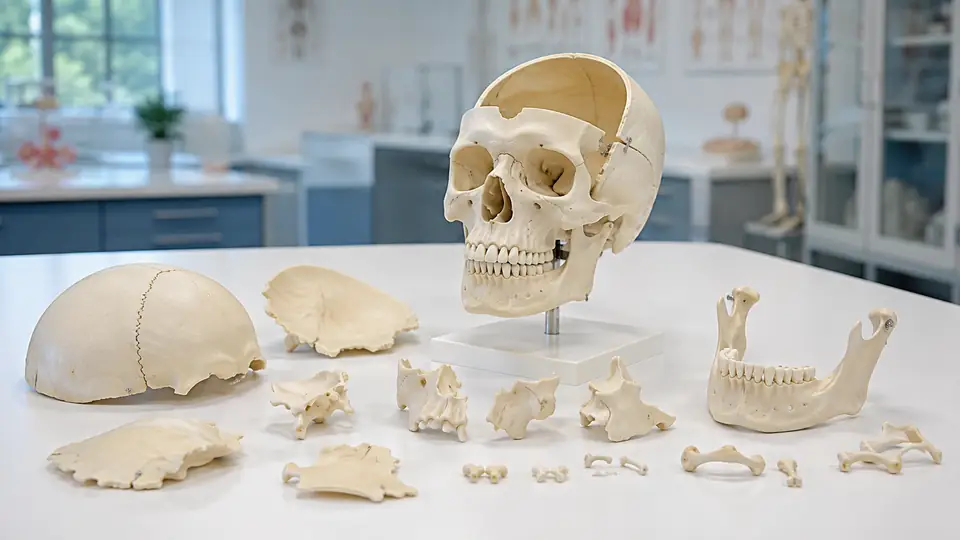
The word disarticulated changes the entire purpose of a skull model. Instead of presenting the skull as one assembled shell, a disarticulated skull model separates its component bones so learners can examine how the pieces fit together. The result is less like a single rigid object on a shelf and more like an anatomy puzzle, with each bone revealing a different part of the skull’s structure.
In practical terms, a disarticulated skull model is an educational replica with removable or separately displayed skull bones. Depending on its design, it may include the frontal, parietal, temporal, occipital, sphenoid, and ethmoid bones, along with the facial bones and mandible. The exact number of pieces varies, so the label does not guarantee that every model separates every bone, which makes the construction details worth checking.
What “Disarticulated” Means in Skull Anatomy
In anatomy, disarticulation means separating bones at the places where they normally meet. In a living skull, many of these connections are immovable or nearly immovable joints called sutures. A model that has been disarticulated represents those relationships by taking the bones apart, rather than permanently locking them into the familiar skull shape.
That distinction is useful during study. A learner looking at an assembled skull may see the coronal suture as a thin line, but a separated frontal and parietal bone show which surfaces create that line. Likewise, removing the temporal bone can expose the neighboring edges of the sphenoid, parietal, and occipital bones, turning a flat boundary into a three-dimensional relationship.
The term does not mean that the model is damaged or intended to represent an injury. It describes the model’s educational arrangement. Much like separating the pieces of a labeled puzzle, disarticulation allows the skull’s construction to be inspected from several angles before the parts are put back together.
How a Disarticulated Skull Model Is Constructed
Most disarticulated skull models use individual molded pieces that correspond broadly to bones or groups of bones. Some pieces are held in place by magnets, pegs, connectors, or shaped contact surfaces. Others are simply designed to rest together. The attachment method affects how easily a learner can remove a bone, how securely the skull stays assembled, and how carefully it must be handled.
Model terminology is not perfectly standardized. One manufacturer might describe a model as a “22-part skull,” while another may count the mandible, small facial bones, or paired bones differently. Some models separate the major cranial bones but keep tiny structures attached, while others divide the skull into a more detailed collection of pieces. A part count is therefore a description of construction, not a universal anatomical standard.
| Model Feature | What It Usually Shows | Why It Helps |
|---|---|---|
| Removable bones | Individual cranial or facial bones that can be taken apart | Shows how neighboring bones meet and overlap |
| Visible sutures | Lines between bones on the assembled skull | Connects surface landmarks with bone boundaries |
| Internal surfaces | Foramina, fossae, ridges, and impressions | Reveals structures hidden inside an assembled skull |
| Separate mandible | The lower jaw displayed independently | Clarifies the temporomandibular joint and dental arch |
| Labels or numbering | Names, codes, or a matching reference chart | Supports identification and self-testing |
Some models also include a transparent section, a removable calvaria, or a braincase that opens. Those features are not the same as complete disarticulation, but they may be combined with it to reveal the cranial cavity, internal base of the skull, or relationship between the braincase and facial skeleton.
Which Skull Structures Can Be Studied?
A disarticulated skull model is especially useful for learning the difference between the neurocranium and the viscerocranium. The neurocranium forms the protective case around the brain, while the viscerocranium includes the facial skeleton. On an assembled skull, those regions blend into one hard contour. Once the pieces are separated, their boundaries become far easier to inspect.
Cranial Bones and Their Neighbors
The frontal bone forms the forehead and contributes to the roof of the orbit. The paired parietal bones make up much of the skull vault, while the occipital bone forms the back and base of the cranium. The temporal bones sit at the sides and contain complex regions associated with the ear. The sphenoid and ethmoid bones are especially important for understanding the skull base, orbit, nasal cavity, and internal cranial landmarks.
Studying these bones separately can answer questions that a diagram may leave fuzzy. Which bone contributes to the medial wall of the orbit? Where does the sphenoid meet the frontal bone? How does the temporal bone connect the side of the skull to its base? With a separated model piece in hand, the answer becomes a matter of tracing edges, surfaces, and neighboring contacts.
Facial Bones, Orbits, and Nasal Structures
Facial bones such as the maxilla, zygomatic, nasal, lacrimal, palatine, and vomer help form the face, orbit, and nasal cavity. Their shapes can be difficult to untangle when they are viewed only from the front. A disarticulated model lets learners rotate one piece, inspect its thin projections, and then return it to the surrounding structure.
The orbits provide a particularly useful exercise because several bones contribute to each eye socket. A student may first identify the large zygomatic and maxilla, then locate smaller contributions from the frontal, sphenoid, ethmoid, and lacrimal bones. That process turns the orbit from a dark hollow into a meeting point made by several distinct bones.
The Mandible and Temporomandibular Joint
The mandible is the only freely movable bone of the skull in ordinary anatomical movement, and it is commonly supplied as a separate piece. Its condylar process meets the temporal bone at the temporomandibular joint. When the mandible is removed from a model, the mandibular fossa and articular tubercle of the temporal bone are easier to locate.
This separation also clarifies the difference between the upper and lower dental arches. The maxilla belongs to the facial skeleton and remains fixed to the cranium, while the mandible forms the lower jaw and moves relative to the temporal bones. That simple contrast is much easier to observe when the lower jaw is not visually fused into the rest of the skull.
Disarticulated Versus Articulated Skull Models
An articulated skull model is assembled into its normal anatomical arrangement. It is useful for studying overall shape, surface landmarks, sutures, foramina, facial proportions, and the general relationship between the cranium and mandible. It is the version that most closely resembles the skull’s intact external form.
A disarticulated model adds another layer by allowing the bones to be examined independently. Neither arrangement is automatically superior. The assembled version is easier to read as a whole, while the separated version is better suited to investigating how the whole is built.
| Feature | Articulated Skull Model | Disarticulated Skull Model |
|---|---|---|
| Bone arrangement | Bones are assembled in anatomical position | Bones can be separated or displayed individually |
| Best for | Overall orientation and surface anatomy | Bone identification and spatial relationships |
| Sutures | Seen as lines between neighboring bones | Can be understood by comparing the actual contacting edges |
| Internal anatomy | May be partly hidden | More accessible when components are removed |
| Handling | Usually simpler to transport and display | Requires organization and careful reassembly |
In classroom teaching, an instructor might use an articulated skull to point out the pterion or foramen magnum, then remove selected bones to show why those landmarks occur where they do. During individual study, a learner can use the same sequence in reverse, first naming a bone and then checking how accurately it fits into the complete skull.
How Students Use a Disarticulated Skull Model
The most productive approach is to combine observation with recall rather than simply reading the labels. Start with a few large bones, such as the frontal, parietal, occipital, and mandible. Identify each from multiple views, then examine what changes when the piece is rotated. The rough curve of the parietal bone, the broad face of the frontal bone, and the distinctive shape of the mandible provide useful visual anchors.
Next, study neighboring relationships. Place the temporal bone beside the sphenoid and occipital bones, trace the base of the skull, and locate openings such as the foramen magnum. Then cover the reference chart and name the bones from their shapes alone. That small pause, with the answer just out of reach, turns the model into a self-testing tool rather than a passive display.
It also helps to compare the physical model with an atlas, labeled diagram, or digital anatomy resource. A physical piece shows thickness and contact surfaces particularly well, while a diagram may label a tiny foramen more clearly. Digital tools can provide rotation or sectional views. Used together, these resources address different parts of the same spatial problem without requiring one tool to do everything.
Common Misunderstandings About These Models
Not every skull advertised as “disarticulated” separates into every individual bone. Some models divide only the major cranial and facial components, while others combine small bones or attach structures that would be difficult to handle independently. Always treat the listed parts and manufacturer instructions as the definitive description of a particular model.
Disarticulation also does not automatically mean that every internal structure is visible. A model may separate the outer bones but still leave the cranial base, nasal cavity, or orbit difficult to inspect. Features such as a removable calvaria, openable braincase, or cutaway section are separate design choices and should not be assumed from the word alone.
Finally, a model is an educational representation, not a perfect substitute for every form of anatomical study. Molded surfaces may simplify thin bones, vary in color, omit small landmarks, or exaggerate features for visibility. Those choices can be helpful for learning, but they should be checked against reliable anatomical references when precise detail matters.
Handling, Cleaning, and Storage
Separate the bones over a clear, stable surface so a small facial component cannot roll under a desk or disappear into a crowded drawer. Keep paired bones organized, and avoid forcing connectors, magnets, or peg fittings if a piece does not seat easily. The fit may depend on a specific orientation, and pressure can damage painted edges or attachment points.
Care instructions depend on the model’s material and finish. Follow the manufacturer’s guidance, especially if the model includes printed labels, painted landmarks, adhesives, magnets, or a stand. In the absence of specific instructions, use gentle handling and avoid solvents, abrasive cleaners, prolonged soaking, and excessive moisture. Store the pieces in a padded, organized container rather than allowing them to knock together in a loose box.
Frequently Asked Questions
Does a disarticulated skull model show real human bone separation?
It represents the bones as separate components, but it is a manufactured teaching aid. The number of pieces, the accuracy of the contact surfaces, and the degree of detail depend on the model’s design.
Are all skull bones removable?
No. Some models separate many major bones, while others combine smaller structures or keep certain parts attached. The listed component count and product documentation provide the most reliable answer for an individual model.
Is a disarticulated skull model useful for beginners?
Yes, provided the learner begins with the larger bones and uses a reference guide. Starting with every small facial bone at once can feel like sorting a handful of nearly identical pieces, so progressive study is usually clearer.
Can the model be used to study sutures?
Yes. The assembled model shows the visible suture lines, while the separated bones help explain which edges form those joints. The model may simplify or exaggerate sutures for visibility, so detailed study should be cross-checked with an anatomical atlas.
The Practical Meaning of Disarticulation
A disarticulated skull model is a skull teaching model designed to separate into identifiable bone components, allowing learners to study both individual anatomy and the way the pieces fit together. Its greatest value lies in that two-step view: first inspect the bone by itself, then rebuild the larger relationships among the cranium, face, orbits, skull base, and mandible.
When the term appears in a model description, look beyond the part count. Check which bones are removable, whether internal surfaces are exposed, how pieces connect, and whether labels or a reference chart are included. Those details reveal what the model can actually demonstrate, turning a general definition into a useful study plan.
