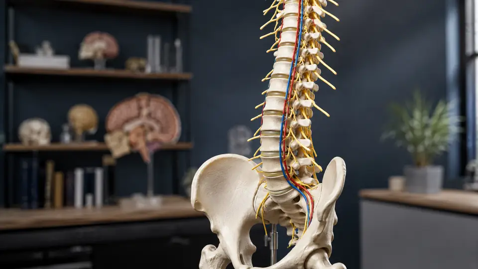
At first glance, a spine model may seem like a neat stack of vertebrae, discs, and joints. Add colored nerves and arteries, however, and the model begins to show the busy anatomy passing around that bony column. The extra structures answer an important question: what travels through the openings and spaces of the spine, and why do those pathways matter?
Nerves and arteries add context, depth, and function to a spine model. They help learners connect the vertebrae with the spinal cord, spinal nerves, and blood vessels that support and serve the nervous system. They also make features such as the vertebral canal and intervertebral foramina easier to understand, although a model is still a simplified teaching aid rather than a complete representation of living anatomy.
What Nerves and Arteries Represent on a Spine Model
On an anatomy model, the structures labeled as nerves usually represent the spinal cord, spinal nerve roots, spinal nerves, or branches leaving the vertebral column. The exact arrangement depends on the model. Some show a central cord inside the vertebral canal, while others emphasize paired spinal nerves emerging through the intervertebral foramina between neighboring vertebrae.
That distinction matters during study. A student looking at a bare vertebral column might identify the foramina as holes between bones. With nerves in place, those openings become passageways with a purpose. The model links a bony landmark to the route taken by a nerve as it leaves the spinal canal and travels toward the trunk or limbs.
Arteries are usually represented by colored vessels placed near the vertebrae, spinal cord, or nerve roots. Depending on the design, a model may show the vertebral arteries traveling upward through the transverse foramina of the cervical vertebrae, or it may include smaller vessels associated with the spinal cord and surrounding tissues. These are not interchangeable structures, so the model’s legend or product documentation should be checked before assigning a name to every colored vessel.
For example, a learner may see a red vessel threading through the openings in the neck vertebrae and associate it with the vertebral arteries. In another model, a smaller vessel near the spinal cord may be intended to suggest arterial supply without showing every individual branch. The color provides a visual cue, but the label provides the anatomical meaning.
How Nerves Make the Spine’s Openings Meaningful
The vertebral canal is formed by the aligned vertebral foramina. It provides a bony passage around the spinal cord, although the cord itself does not extend through the entire length of the adult vertebral column. The spinal cord typically ends in the upper lumbar region, while nerve roots continue downward within the canal as the cauda equina before exiting at appropriate levels.
A spine model with a cord and nerve roots can make that arrangement easier to see than a paragraph alone. A learner can follow the cord inside the canal, observe the lower bundle of nerve roots, and then trace individual nerves toward the intervertebral foramina. That spatial relationship is difficult to capture in a single flat image, especially when the model can be viewed from the back, side, and front.
The paired spinal nerves are formed from sensory and motor roots that connect with the spinal cord. After leaving the vertebral column, they divide into branches that serve different regions of the body. A model may simplify these branches or omit them entirely, but even the short segment shown beside each vertebra can clarify that the nerves are not embedded randomly in the bone.
Consider a classroom demonstration of the cervical spine. Without nerves, the transverse processes, pedicles, and foramina may look like a complicated row of ridges and gaps. With a nerve pathway added, the instructor can point from the spinal cord to the nerve root and then to the opening between vertebrae, turning a three-dimensional route into something the group can follow with its eyes.
Spinal Nerves Versus The Spinal Cord
These terms are related but not identical. The spinal cord is the long central structure within the vertebral canal. Spinal nerves are paired nerves that connect with the cord and leave the canal through openings in the spine. Some models use one continuous colored piece for the cord and separate pieces for the nerve roots, while others use a simplified combined representation.
This is one reason a spine model should not be read by color alone. A bright yellow structure may indicate nerves, but it may not show every root, branch, or connection exactly as it appears in the body. If the model does not clearly distinguish the cord, roots, and spinal nerves, an anatomy atlas or labeled diagram can fill in the missing detail.
How Arteries Add Circulation and Orientation
Arteries give a spine model another layer of meaning. Bone establishes the framework, nerves show communication, and blood vessels suggest the circulation that supplies the spinal cord, nerve roots, vertebrae, and nearby tissues. This helps learners understand that the spine is not an isolated stack of mechanical parts.
The vertebral arteries are a particularly useful feature when they are shown. They travel through the transverse foramina of several cervical vertebrae and continue toward the cranial cavity. Seeing a vessel pass through those repeated openings gives the transverse foramina a clear anatomical role rather than leaving them as decorative holes on the sides of the neck vertebrae.
Models may also show arteries associated with the spinal cord and nerve roots. In the body, blood supply to the spinal cord involves longitudinal vessels and reinforcing branches entering at different levels. The exact pattern is complex and variable, so a teaching model may display only selected vessels or use a schematic route instead of attempting a complete vascular map.
A learner studying the thoracic and lumbar regions may notice that the vessels are thinner and less visually dominant than the vertebrae. That is intentional in many models. The bones remain the main subject, while the vessels provide orientation and introduce the idea that smaller branches reach structures hidden within the canal and surrounding tissues.
What These Features Help You Study
Nerves and arteries are most valuable when they help explain relationships, not when they are treated as decoration. A useful study session might begin with the vertebrae alone, followed by identification of the canal, foramina, nerve pathways, and vessels. Each added structure answers a different question about the same region.
- Intervertebral foramina: Nerves show why these openings matter and where spinal nerves leave the vertebral column.
- Vertebral canal: The spinal cord or nerve-root bundle helps define the protected space formed by the vertebrae.
- Cervical transverse foramina: Vertebral arteries, when included, demonstrate the passage of vessels through the neck vertebrae.
- Regional orientation: Nerves and vessels help distinguish anterior, posterior, lateral, cervical, thoracic, and lumbar relationships.
- Clinical terminology: The model can support explanations of nerve roots, spinal nerves, vascular supply, and narrowing of anatomical passageways.
During individual study, a learner might cover the labels and trace one nerve from the cord to the side of the spine, then identify the vertebra and opening involved. In a group setting, someone can rotate the model while another person describes what is visible from the posterior or lateral view. Those small tasks turn a static object into a spatial quiz.
The added structures are also useful for comparing regions. Cervical vertebrae have transverse foramina, while typical thoracic and lumbar vertebrae have different shapes and landmarks. If a model shows vertebral arteries only in the cervical region, that visual contrast reinforces the fact that not every opening in the spine carries the same structure.
What A Nerve-And-Artery Spine Model Does Not Show
Even a detailed model is selective. It may enlarge the nerves and vessels so they remain visible beside the much larger vertebrae. It may also use bright colors, smooth surfaces, or flexible tubing that make the structures easier to identify but do not reproduce their natural appearance.
A model may leave out small arterial branches, venous structures, connective tissue, membranes, fat, and the fine anatomy of individual nerve roots. It may show the spinal cord as a simple cylinder rather than representing its changing shape, enlargements, coverings, or internal organization. These omissions are not necessarily flaws. They allow the major relationships to remain visible from across a room or on a crowded study table.
The model also cannot show movement, pulsation, tissue texture, or the way structures change position with posture. A flexible spine may demonstrate bending, but its attached nerves and arteries may not move exactly as living tissues do. If a model’s cord or vessels are fixed in place, treat the movement as a broad anatomical illustration rather than a precise simulation.
For instance, rotating a spine model to show a narrowed opening can demonstrate the location of the foramen. It cannot determine what a particular person’s nerves or blood vessels look like, and it cannot be used to diagnose a condition. The teaching value lies in understanding normal structure and spatial relationships.
How To Read The Colors And Labels
Color conventions are common, but they are not universal. Yellow often suggests nerves, red commonly suggests arteries, and blue may suggest veins, yet manufacturers may use different palettes or apply colors for contrast rather than strict anatomical convention. The printed key, numbered labels, or accompanying guide should take priority over assumptions.
Start by identifying the model’s central reference points. Find the vertebral bodies, posterior elements, vertebral canal, and intervertebral foramina. Then determine whether the colored structures represent the spinal cord, nerve roots, spinal nerves, vertebral arteries, or a more generalized set of pathways.
A practical approach is to ask three questions for each added structure:
- What is this structure called?
- Where does it travel in relation to the vertebrae?
- What opening, surface, or region helps explain its position?
Suppose the label identifies a vertebral artery. The next step is not simply memorizing the name. Trace the vessel through the cervical transverse foramina and compare that path with the location of the spinal cord and spinal nerves. The relationship is the lesson, and the label is only the starting point.
Nerves And Arteries Compared With A Bare Spine Model
| Feature | Bare Spine Model | Spine Model With Nerves And Arteries |
|---|---|---|
| Main emphasis | Vertebrae, discs, joints, and bony landmarks | Bony anatomy plus neural and vascular relationships |
| Foramina | Visible as openings in the bone | Shown as passageways for selected nerves or vessels |
| Vertebral canal | Demonstrates the surrounding bony space | Provides context for the spinal cord and nerve roots |
| Cervical transverse foramina | Shows their shape and location | May show the route of the vertebral arteries |
| Study focus | Osteology, articulation, and spinal alignment | Osteology combined with neuroanatomical and vascular orientation |
Neither design is automatically better for every lesson. A bare model may be less visually crowded when the goal is to compare vertebral landmarks or demonstrate spinal movement. A model with nerves and arteries is more informative when the lesson centers on what passes through and around those landmarks.
The difference becomes obvious when studying the intervertebral foramen. On a bare model, the opening is easy to inspect. On a model with a nerve pathway, the same opening becomes part of a route that can be traced from the canal outward. One model isolates the bone, while the other places the bone in a larger anatomical system.
Using These Features For Active Study
Rather than reading every label in sequence, use the model to ask location-based questions. Cover the labels and identify the structure by its position. Trace a vessel from one cervical level to the next. Point to the canal, then name what it contains and how that changes in the lower spine.
A second useful exercise is comparison. Look at a cervical, thoracic, and lumbar region and ask which nerve or vessel features are shown in each area. Then check the model guide or an atlas. This approach exposes oversimplification quickly, especially when a model shows only representative vessels rather than every anatomical branch.
For a small study group, one person can choose a structure while another rotates the model to find it from a different angle. A third person can explain what lies anterior, posterior, or lateral to it. The exercise resembles a verbal map, with the spine serving as the fixed terrain and the nerves and arteries marking important routes.
Pairing the model with a labeled illustration is especially useful for details the physical model cannot display. A diagram can show the roots, branches, coverings, and vascular connections in greater detail, while the three-dimensional model clarifies depth and orientation. Used together, the resources answer different parts of the same anatomy question.
Common Misunderstandings About The Added Structures
Every Colored Vessel Is Not Necessarily A Complete Artery
A red tube may represent an artery, but it may also be a simplified pathway or selected example. Read the key before assuming that the model depicts every arterial branch. Anatomy models often prioritize visibility over completeness.
Nerves Do Not Fill Every Opening In The Same Way
The presence of a nerve near a foramen does not mean that every opening contains an identical structure. Spinal levels differ, and the model may show representative nerves rather than a fully detailed arrangement. The shape and region of the vertebra still matter.
The Model Is Not A Diagnostic Device
A spine model can help explain where a nerve or artery is located, but it cannot show an individual’s anatomy or confirm a medical condition. Its purpose is education, demonstration, and spatial reference.
Final Takeaway
Nerves and arteries turn a spine model from a study of bones into a broader map of the structures that pass through and around the vertebral column. Nerves clarify the spinal cord, nerve roots, spinal nerves, and intervertebral foramina. Arteries add circulation and help explain pathways such as the vertebral arteries through the cervical transverse foramina.
The most useful way to interpret these features is relationally. Identify the vessel or nerve, trace its route, connect it to the surrounding bony landmark, and confirm the model’s labels before drawing detailed conclusions. That approach keeps the model accurate in the ways that matter most while respecting what every simplified teaching aid leaves out.
