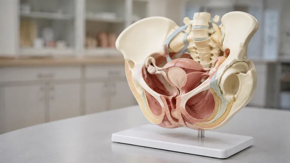
The female pelvis is a compact maze of bone, muscle, ligaments, nerves, and closely arranged organs. In a lecture, those relationships can blur into a crowded slide. Place a dimensional model on the desk, rotate it toward the sacrum, and suddenly the pelvic outlet, pubic symphysis, and surrounding structures make spatial sense. Buyers seek these models for anatomy study, midwifery education, classroom demonstrations, artistic reference, and clearer patient conversations.
The main purchasing mistake is focusing on appearance without checking what the model actually shows. A flexible skeletal pelvis teaches different lessons from a fixed pelvic-floor model, while removable organs are far more useful for tracing spatial relationships. Scale, articulation, color coding, included structures, and durability matter most. The Female Pelvic Muscle and Perineum Model leads this list for its broad anatomical coverage, life-size format, movable coccyx, and teaching manual, but learners who need removable organs or movable hip joints have compelling alternatives.
Priorities vary sharply. A student studying at a cramped kitchen table may appreciate a palm-friendly miniature, while an instructor facing a full classroom needs a larger, visually clear display. The strongest choice is the one that matches the anatomy being taught and the distance from which it will be viewed.
Quick Answer
Best overall: The Female Pelvic Muscle and Perineum Model combines bones, pelvic-floor muscles, ligaments, nerves, and perineal anatomy in a life-size teaching format. The Female Pelvis and Perineum 6-Part Model is better for learners who want removable organs, while the JNMFTD Lumbar Spine and Female Pelvis Model provides the broadest view of the pelvis, lumbar spine, hip joints, muscles, and nerves. For portable skeletal study, the XUYUH Mini Female Pelvis Model is the compact pick.
For a quick side-by-side view, the table highlights the structural differences that matter most.
Best Female Pelvis Models Comparison Table
| Product | Best For | Scale or Size | Key Feature |
|---|---|---|---|
| Female Pelvic Muscle and Perineum Model | Overall anatomy teaching | Life-size, 10.6 x 7.8 x 7.1 inches | Muscles, ligaments, nerves, perineum, and manual |
| Female Pelvis and Perineum 6-Part Model | Organ relationships | Life-size format | Removable uterus and bladder with magnetic mounting |
| Merinden Movable Female Pelvis Model | Hip mechanics | Life-size | Movable femur heads, joints, and elastic connections |
| Evotech Flexible Female Pelvis Model | Midwifery and skeletal study | About 9.6 x 6.7 x 6.7 inches | Flexible, separable bones |
| Benilev Female Pelvis Muscle Model | Fixed pelvic-floor display | 10.6 x 7.8 x 7.1 inches | Color-coded muscles, ligaments, nerves, and perineum |
| JNMFTD Lumbar Spine and Female Pelvis Model | Broad regional anatomy | 1:1 life-size | Lumbar spine, nerves, muscles, and movable femur heads |
| LYOU Life-Size Flexible Female Pelvis Model | Flexible classroom demonstrations | About 9.6 x 6.7 x 6.7 inches | Flexible bones and rubber pubic symphysis |
| XUYUH Mini Female Pelvis Model | Compact study | About 5.9 x 5.1 x 2.75 inches | Small, flexible, and portable |
| 3B Scientific A61 Female Pelvis Model | Premium skeletal reference | Natural-size format | Three-part female pelvic skeleton |
The detailed reviews below show where each model earns its place and which anatomical lessons it handles best.
1. Female Pelvic Muscle and Perineum Model: Best Overall

This life-size model earns the top position by showing far more than bare pelvic bones. Its visible pelvic-floor muscles, ligaments, nerves, and perineal structures create a layered teaching surface with splashes of color separating adjacent anatomy. The pelvic assembly includes hip bones, the sacrum, a movable coccyx, and two lumbar vertebrae. Reproductive and rectal insertions add context, helping a learner see how soft tissues occupy the bony basin rather than floating in isolation on a page.
At approximately 10.6 by 7.8 by 7.1 inches, it is large enough for desk-side instruction without becoming an awkward lump to store. Picture an instructor turning it toward a small group and pointing from the lumbar region down through the pelvic floor. The movement follows a visible anatomical path, which is much clearer than hopping between separate illustrations. Color differentiation and the included teaching manual further support structured lessons, while washable PVC suits repeated classroom handling.
Pros
- Displays pelvic bones, muscles, ligaments, nerves, and perineal anatomy
- Life-size proportions support realistic spatial study
- Includes a movable coccyx and two lumbar vertebrae
- Color differentiation makes neighboring structures easier to distinguish
- Includes a teaching manual for guided reference
Cons
- Less interactive than models with multiple removable organs
- Dense anatomy may feel visually busy for an introductory skeletal lesson
This is the strongest all-purpose choice for medical students, pelvic-health educators, nursing programs, and clinicians who need to explain several tissue layers with one display. Buyers focused solely on pelvic bone mechanics can save space with a skeletal model. For a richer discussion of the pelvic floor and perineum, however, this colorful life-size format provides much more to point at, trace, and discuss.
To buy this product, click here.
2. Female Pelvis and Perineum 6-Part Model: Best for Removable Organs

Removable anatomy changes the lesson from passive viewing to spatial problem-solving. This six-part model depicts the pelvic floor and genital tract alongside the uterus, vagina, ovaries, bladder, and rectum. The uterus and bladder can be removed, allowing students to inspect structures that would otherwise disappear behind neighboring organs. Magnetic mounting helps return the components to their intended positions without a fiddly balancing act.
That arrangement is particularly helpful during a small-group tutorial. A student can lift away the bladder, peer into the exposed cavity, and then replace it to understand how closely the organs sit together. The model also includes hip bones, a sacral region with a movable coccyx, and two lumbar vertebrae. Its PVC construction can be wiped clean after frequent handling, which matters in a lab where the same model may pass through a dozen pairs of hands before lunch.
Pros
- Six-part design supports interactive anatomical study
- Removable uterus and bladder reveal deeper relationships
- Magnetic mounting simplifies component placement
- Shows pelvic-floor and perineal structures alongside organs
- Washable PVC construction suits recurring educational use
Cons
- Loose components require more careful storage than a fixed model
- Reassembly adds complexity for quick room-wide demonstrations
Students learning organ position, pelvic fascial levels, or the relationship between the uterus, bladder, and rectum will gain the most from this model. It also suits educators who like to ask learners to remove, identify, and replace structures during practical sessions. A fixed model is faster to carry between rooms, but it cannot provide the same revealing look beneath the visible surface.
To buy this product, click here.
3. Merinden Movable Female Pelvis Model: Best for Hip Mechanics

The Merinden model shifts attention from internal organs to motion at the pelvis and hips. It uses elastic connections and includes two movable femur heads and joints, creating a hands-on way to explore how the pelvic skeleton meets the upper femur. For orthopedic instruction, gait-related anatomy, or a basic demonstration of hip articulation, that movement carries more teaching value than painted soft tissues.
Imagine explaining the acetabulum with the femoral head already seated beside it. Rather than tracing a tiny circular socket in a book, an instructor can move the joint and show the relationship from several angles. Its life-size construction preserves realistic proportions, while the flexible assembly invites supervised handling in nursing schools, biology classrooms, and professional presentations. The design also works for patient education where a simple moving joint communicates more cleanly than a dense wall chart.
Pros
- Life-size pelvis provides realistic skeletal proportions
- Two movable femur heads show the pelvis-to-femur relationship
- Elastic mounting permits flexible, interactive demonstrations
- Useful for orthopedic, obstetric, and general anatomy education
- Durable construction is intended for frequent handling
Cons
- Does not provide the soft-tissue detail of pelvic-floor models
- Flexible connections are less rigid than a fixed display assembly
This is a persuasive choice for learners who need movement rather than removable organs. It works especially well on a demonstration table where the instructor and student can rotate the hip joints together. Buyers studying nerves, reproductive organs, or layered pelvic-floor musculature should choose a more detailed regional model, but the Merinden keeps bone and joint mechanics refreshingly clear.
To buy this product, click here.
4. Evotech Flexible Female Pelvis Model: Best Value

For focused study of female pelvic bones, the Evotech strips away visual clutter. The model is cast from a selected natural female pelvis and presented at life size, approximately 9.6 by 6.7 by 6.7 inches. Its bones are mounted with elastic cord, allowing the joints to separate slightly so learners can inspect meeting surfaces and gaps that remain hidden on a rigid specimen.
The pubic symphysis uses rubber to emphasize the cartilaginous connection, a small but useful detail during obstetric or midwifery lessons. Set it beside a notebook and the model becomes easy to rotate while sketching the inlet, outlet, sacrum, and pubic arch. It can also be disassembled, adding another layer of interaction for supervised demonstrations. PVC bones keep maintenance simple, although this skeletal approach deliberately leaves out organs and muscle layers.
Pros
- Life-size model is cast from a selected natural female pelvis
- Elastic mounting permits flexible joint demonstrations
- Can be disassembled for closer inspection
- Rubber pubic symphysis highlights an important pelvic connection
- Focused skeletal design is easy to read without soft-tissue clutter
Cons
- Does not depict reproductive organs, nerves, or pelvic-floor muscles
- Disassembled parts need attentive handling and storage
Midwifery students, anatomy beginners, and instructors teaching pelvic dimensions may prefer this straightforward format over a crowded multicolored model. It is particularly practical where learners need to hold and reposition the bones. Those preparing a detailed pelvic-floor lesson will require a second resource, but for tactile skeletal anatomy, the Evotech provides a clean and flexible foundation.
To buy this product, click here.
5. Benilev Female Pelvis Muscle Model: Best Fixed Pelvic-Floor Display

Some teaching sessions benefit from a model that stays assembled and ready to point at. This Benilev display is non-detachable, keeping its pelvic bones, pelvic-floor muscles, ligaments, nerves, and perineum together as a single reference. Separate colors mark neighboring structures, producing a vivid map for anatomy review or a consultation-room explanation where loose components would become a distraction.
The model measures roughly 10.6 by 7.8 by 7.1 inches, a substantial desktop footprint that gives small structures room to remain visible. During a study session, learners can circle it, compare anterior and posterior landmarks, and follow colored tissues across the pelvic floor without pausing to rebuild anything. The PVC surface is washable and intended for reuse, making the model appropriate for classrooms, laboratories, offices, and dedicated study spaces.
Pros
- Displays pelvic-floor muscles, ligaments, nerves, and perineum
- Color-coded structures improve visual separation
- Fixed construction keeps the anatomy ready for immediate display
- Desktop dimensions support close individual or small-group viewing
- Washable PVC is convenient for shared learning environments
Cons
- Non-detachable anatomy prevents inspection beneath overlapping structures
- Less useful than articulated models for demonstrating joint movement
This model fits educators who want a stable visual aid on a shelf or consultation desk. It is also a sensible choice for students who dislike the clatter and reassembly of multipart models. Learners who need to remove the bladder or uterus should select the six-part option, while those focused on hip motion will find more value in movable femur heads. Here, the appeal is immediate, colorful pelvic-floor visibility.
To buy this product, click here.
6. JNMFTD Lumbar Spine and Female Pelvis Model: Best Detailed Regional Model

The JNMFTD broadens the field of view above and below the pelvis. Its 1:1 assembly includes the lumbar spine, pelvis, femurs, hip joints, muscles, and nerves, providing a regional reference rather than an isolated bony ring. That expanded anatomy helps connect lumbar structures to the pelvis and then into the proximal femurs, a useful sequence for orthopedic education and general musculoskeletal study.
Color-marked features and an included product manual support identification, while movable femur heads bring mechanical context to the display. The model comes with a sturdy base and can be removed from it, allowing a teacher to switch between a steady upright presentation and closer tabletop inspection. Picture a classroom discussion moving from lumbar nerves to the hip joint in one continuous sweep. The physical continuity keeps the explanation grounded and visible.
Pros
- Combines lumbar spine, female pelvis, femurs, muscles, and nerves
- Movable femur heads support hip-joint demonstrations
- Color marking distinguishes selected anatomical features
- Includes a product manual for reference
- Removable sturdy base supports display and closer handling
Cons
- Larger regional scope requires more storage space than pelvis-only models
- Broad musculoskeletal coverage is less focused on reproductive organs and perineal layers
Educators who routinely move between lumbar, pelvic, and hip anatomy will appreciate having those regions united on one base. It also makes a striking office display for patient-friendly skeletal explanations. A compact pelvis is easier to slip into a cabinet, and a removable-organ model offers deeper visceral study. The JNMFTD earns its place by showing how the larger region connects, rather than treating the pelvis as an isolated island.
To buy this product, click here.
7. LYOU Life-Size Flexible Female Pelvis Model: Best for Classroom Handling

The LYOU model favors a simple, tactile approach. At about 9.6 by 6.7 by 6.7 inches, it provides life-size pelvic proportions without surrounding organs or a tall display base. Its bones are flexibly connected and can be removed, allowing students to inspect the pelvic skeleton from angles that a rigid assembly may resist.
A rubber pubic symphysis adds material contrast at the front of the pelvis, while washable PVC makes the model suitable for a busy anatomy room. Think of a lab station where pairs of students identify landmarks, rotate the sacral region, and then pass the model along. The uncluttered bone surfaces keep that exercise focused. It also suits obstetric, gynecologic, and orthopedic explanations where the skeletal framework is the central subject.
Pros
- Life-size dimensions support realistic skeletal study
- Flexible bones permit changes in viewing angle
- Removable construction allows closer inspection
- Rubber pubic symphysis emphasizes the anterior joint
- Washable PVC works well in shared classrooms and offices
Cons
- Provides no pelvic-floor muscles or internal organs
- Flexible mounting offers less display rigidity than a fixed skeleton
The LYOU is best for instructors and students who expect frequent handling and want the anatomy kept visually simple. It competes closely with the Evotech, sharing a similar life-size, flexible concept. Choose this type for bony landmarks and pelvic shape, rather than detailed soft-tissue relationships. Its uncomplicated form is precisely what makes it approachable during introductory practical work.
To buy this product, click here.
8. XUYUH Mini Female Pelvis Model: Best Compact Option

Space disappears quickly on a student desk once the laptop, notes, highlighters, and coffee mug arrive. The XUYUH addresses that familiar squeeze with a simulated-size pelvis measuring approximately 5.9 by 5.1 by 2.75 inches. It is much smaller than the life-size models here, making it easier to store in a drawer or carry between home, class, and study sessions.
The model retains flexible bone connections and uses rubber at the pubic symphysis. Its PVC construction can be washed, a practical detail for a portable object that may spend time in a backpack. Close-up viewing is necessary because of the reduced size, but the miniature form still offers a physical reference for pelvic orientation and major skeletal relationships. For one learner turning it under a desk lamp, that portability may matter more than room-wide visibility.
Pros
- Compact dimensions make storage and transport easier
- Flexible construction allows varied viewing angles
- Rubber pubic symphysis provides useful material contrast
- Washable PVC is practical for regular handling
- Well suited to personal desks and individual study
Cons
- Reduced scale makes small landmarks harder to view from a distance
- Does not include internal organs, muscles, nerves, or ligaments
This miniature is aimed at individual learners, traveling educators, and buyers with tight storage limits. It is less convincing for a classroom where students in the back row need to see the pelvic inlet clearly. A life-size option offers better landmark visibility, but it cannot match the convenience of a model that slips neatly into a bag after the study session ends.
To buy this product, click here.
9. 3B Scientific A61 Female Pelvis Model: Best Premium Skeletal Reference
The 3B Scientific A61 is a focused three-part representation of the female pelvic skeleton. Its natural-size format supports close study of pelvic shape and bony relationships without covering the surface in soft-tissue colors. For educators who prefer a restrained skeletal reference, this type of model fits neatly beside textbooks, radiographic examples, and other osteology materials.
Its value becomes apparent during a lesson centered on major landmarks rather than organ placement. An instructor can orient the pelvis, identify the sacral and hip-bone regions, and discuss the overall inlet and outlet without bright muscle layers pulling the eye elsewhere. The three-part construction also provides more physical context than a flat skeletal plate. It is a premium-leaning choice for established teaching rooms where a dedicated female pelvic skeleton will see repeated use.
Pros
- Natural-size format supports realistic skeletal orientation
- Three-part design provides focused female pelvic anatomy
- Uncluttered surfaces keep attention on bony structures
- Suitable for osteology, obstetric, and general anatomy instruction
- Established educational-model format suits recurring demonstrations
Cons
- Does not show pelvic-floor muscles, nerves, or reproductive organs
- Offers less movement-focused detail than models with movable femur heads
Choose the A61 for a dedicated skeletal reference where clarity and natural proportions outweigh removable organs or bright tissue coding. Students seeking the widest range of anatomy from one purchase will get more visual content from the top overall pick. In a classroom already equipped with organ models, however, this cleaner pelvic skeleton can fill the bony-anatomy role without unnecessary overlap.
To buy this product, click here.
How to Choose a Female Pelvis Model
Start With the Anatomy You Need to See
A bare skeletal pelvis is ideal for studying the pelvic inlet, outlet, pubic arch, sacrum, and major bony landmarks. It keeps the view clean, much like clearing every extra paper from a desk before tackling one difficult diagram. Pelvic-floor models serve a different purpose. Their muscles, ligaments, nerves, and perineal structures help learners understand how soft tissues span and occupy the skeletal framework.
Models with reproductive organs, bladder, and rectum are more appropriate for spatial organ study. Removable components add another advantage by exposing structures hidden inside an assembled pelvis. Match the purchase to the upcoming lesson rather than choosing the model with the greatest number of visible colors.
Compare Life-Size and Miniature Formats
Life-size models provide easier landmark recognition and more realistic spatial relationships. They work well at a lab bench, consultation desk, or small-group teaching table. Larger structures also give an instructor enough room to place a pointer precisely, avoiding the awkward tap-tap-tap of searching for a tiny landmark.
Mini models are easier to carry and store, but their small surfaces demand close viewing. They suit individual study better than lectures. Before buying, measure the intended shelf or cabinet and consider how far the audience will sit from the model.
Choose the Right Type of Movement
“Flexible” can describe several useful features. Elastic-mounted pelvic bones may separate slightly or change angle, making joints and meeting surfaces easier to inspect. Movable femur heads demonstrate the hip connection and are especially relevant for musculoskeletal teaching. A movable coccyx offers a more focused feature at the lower sacral region.
Movement creates a lively hands-on lesson, though flexible models may feel less steady on display than rigid versions. A fixed model is often preferable for quick pointing during a consultation. Articulated designs shine during practical sessions where learners are expected to manipulate the anatomy.
Decide Whether Removable Parts Are Worth the Extra Handling
Removable organs can reveal layered anatomy in a way fixed displays cannot. Lift out the bladder, and the space behind it becomes immediately visible. Replace it, and the close packing of the pelvic organs returns. That simple remove-and-rebuild sequence can turn a confusing relationship into a memorable one.
Multipart models require organized storage. Small components can be misplaced, and students need time to learn the correct orientation. Magnetic mounting is useful because it helps guide pieces into position, while fixed anatomy remains the easier choice for busy offices and fast demonstrations.
Look for Clear Color Coding and Reference Material
Color coding helps distinguish adjacent muscles, ligaments, and nerves, particularly in the crowded pelvic floor. More color is not automatically better. The useful question is whether the colors separate structures cleanly and support the lesson at hand. A skeletal model may need little or no color beyond contrasting cartilage.
An included manual can help learners identify unfamiliar features and plan a logical study sequence. Product documentation varies in depth, so buyers using a model for formal study should still keep an anatomy atlas or course material nearby. A physical model is a visualization aid rather than a replacement for a complete educational reference.
Think About Durability, Cleaning, and Storage
PVC is common across these selections because it is durable and easy to wipe clean. That matters in shared classrooms, where fingerprints and dust accumulate quickly. Flexible cords, magnets, removable pieces, and movable joints introduce additional handling points, so such models benefit from careful supervision and a dedicated storage container.
Check whether a base is included and whether the model can be removed from it. Bases improve visibility and stability on a shelf, while base-free skeletal models are often easier to rotate in the hands. The better arrangement depends on whether the model will spend more time being displayed or passed around.
Best Female Pelvis Models FAQ
Is a Life-Size Female Pelvis Model Better for Students?
Life-size models generally make landmarks and spatial relationships easier to see. They are well suited to lab benches, small groups, and repeated anatomy practice. A miniature can still work for personal review, especially where portability and storage are more important than realistic scale.
Should I Choose a Skeletal Model or a Pelvic-Floor Muscle Model?
Choose a skeletal model for bony landmarks, pelvic shape, joint relationships, and basic obstetric dimensions. Select a pelvic-floor model for muscles, ligaments, nerves, perineal anatomy, and soft-tissue orientation. Learners covering both subjects may prefer a detailed combined model or use separate focused references.
Why Are Removable Organs Useful?
Removable organs expose structures and spaces hidden within the assembled pelvis. They help students visualize how the uterus, bladder, and rectum relate to one another. The trade-off is extra reassembly and storage, so fixed models can be more convenient for quick demonstrations.
What Does a Flexible Female Pelvis Model Demonstrate?
Flexible mounting may allow the bones to separate slightly, change angle, or be removed for inspection. This helps demonstrate joints and pelvic structure. Models with movable femur heads add hip articulation, while a movable coccyx provides movement at a more localized structure.
Are Female Pelvis Models Suitable for Patient Education?
They can support clear, general anatomical explanations by giving viewers a visible three-dimensional reference. Simpler models may be less intimidating for broad skeletal discussions, while color-coded or removable-organ models can clarify specific structures. They remain educational aids and do not replace individualized medical evaluation or advice.
How Should an Anatomy Model Be Cleaned?
Follow the manufacturer’s care guidance. PVC models are commonly wiped with a soft damp cloth and mild cleaning solution, then dried before storage. Avoid aggressive solvents, rough scrubbing, prolonged soaking, and force around cords, magnets, or movable joints.
Do Color-Coded Models Improve Anatomy Study?
Clear color separation can make adjacent muscles, nerves, ligaments, and organs easier to identify. It is most helpful in anatomically dense regions such as the pelvic floor. Color should supplement accurate course materials, since schemes may differ among models and textbooks.
Which Model Is Best for Midwifery Education?
A life-size flexible skeletal pelvis is useful for pelvic shape and bony relationships. A model with pelvic-floor tissues or removable organs provides additional context for soft-tissue and visceral anatomy. Many midwifery learners will benefit most from selecting the model that matches the immediate module rather than seeking one display to cover every topic.
Final Verdict
The Female Pelvic Muscle and Perineum Model is the strongest overall option among these nine picks. Its life-size design combines pelvic bones with muscles, ligaments, nerves, perineal anatomy, a movable coccyx, and two lumbar vertebrae. That broad coverage makes it adaptable for student revision, small-group instruction, and visual explanations. It has enough detail to keep advanced lessons interesting without requiring a box of loose pieces after every session.
Different needs point toward different winners. The Female Pelvis and Perineum 6-Part Model is the better choice for removable-organ study, while the JNMFTD model connects the pelvis to the lumbar spine, femurs, nerves, and muscles. Buyers seeking straightforward skeletal value can look to the flexible Evotech, and the tiny XUYUH is easier to carry between a crowded desk and class. Before purchasing, decide whether the next lesson centers on bones, movement, pelvic-floor tissues, or organ relationships. That single decision will narrow these nine models faster than any long feature list.
