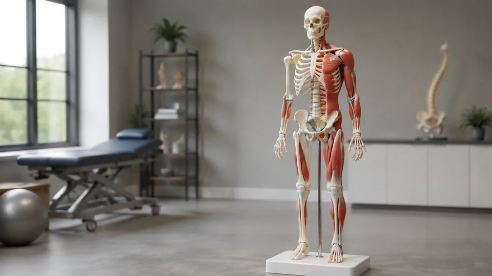
The shoulder looks simple on a page until you try to explain how the humerus, scapula, and surrounding ligaments move together. That is where a three-dimensional skeleton earns its floor space. Models with muscle markings and ligaments let students trace attachment points, teachers demonstrate joint mechanics, and clinicians give patients a clearer visual reference than a dense illustration can provide.
The products in this category vary more than their pale PVC bones might suggest. Some are life-size skeletons with rolling stands, detachable limbs, numbered bones, and painted muscle origins and insertions. Others concentrate on a hand, foot, or group of major joints, trading whole-body context for close, tactile detail. Buyers often focus on height and overlook articulation, fixed spines, removable components, labeling, storage, and the structures actually represented. Different settings demand different features, but the 70.8-inch Human Anatomy Model leads this selection with its broad mix of muscle markings, flexible joint ligaments, nervous-system detail, movable parts, and practical rolling stand.
For a fast look at the strongest choices and their intended uses, start with the quick recommendations and comparison table below.
Quick Answer
Best overall: The 70.8-Inch Human Anatomy Model with Muscle Insertions and Ligaments offers the most versatile feature set for general anatomy instruction. Best compact option: The Axis Scientific Mini Human Skeleton fits desks and smaller study rooms while retaining painted attachment points. Best for joint mechanics: The six-piece Human Joint Anatomy Model Set gives learners separate shoulder, elbow, hip, knee, hand, and foot models that can be handled around a table. For highly detailed foot and ankle study, the premium 3B Scientific M34/1 provides far more regional anatomy than a basic foot skeleton.
Best Skeleton Models With Muscles and Ligaments Comparison Table
| Product | Best For | Size or Scope | Key Anatomical Features |
|---|---|---|---|
| 70.8-Inch Human Anatomy Model | Best overall | 70.8-inch full body | Muscle attachment markings, joint ligaments, numbered bones, nerves, and carotid artery |
| Medical Life-Size Skeleton Replica | Medical and nursing instruction | 71-inch full body | Spinal nerves, muscle origin and insertion points, and joint ligaments |
| Breesky Skeleton with Ligaments | Classroom demonstrations | 70.8-inch full body | Flexible joint ligaments, muscle markings, numbered bones, and detachable limbs |
| Life-Size Skeleton Anatomy Model | Protected storage | 70.8-inch full body | Muscle markings, joint ligaments, nervous-system detail, and dust cover |
| Six-Piece Human Joint Model Set | Joint mechanics | Six life-size joint regions | Articulated ligaments across the shoulder, elbow, hip, knee, hand, and foot |
| Numbered Hand Skeleton Model | Hand anatomy | Seven-piece regional model | Bones, muscles, ligaments, nerves, arteries, and numbering |
| Foot Skeleton Model | Budget foot study | Life-size regional model | Bones, muscles, ligaments, nerves, and blood vessels |
| 3B Scientific M34/1 Foot Skeleton | Detailed foot and ankle study | Foot and lower leg | Muscles, tendons, ligaments, nerves, vessels, tendon sheaths, and removable parts |
| Axis Scientific Mini Human Skeleton | Compact study spaces | 31-inch full body | Painted and numbered muscle origins and insertions with articulated joints |
The full reviews below explain where each model fits, from lecture halls with rolling skeletons to quiet desk study with a hand or foot under a bright task lamp.
1. 70.8-Inch Human Anatomy Model With Muscle Insertions and Ligaments: Best Overall
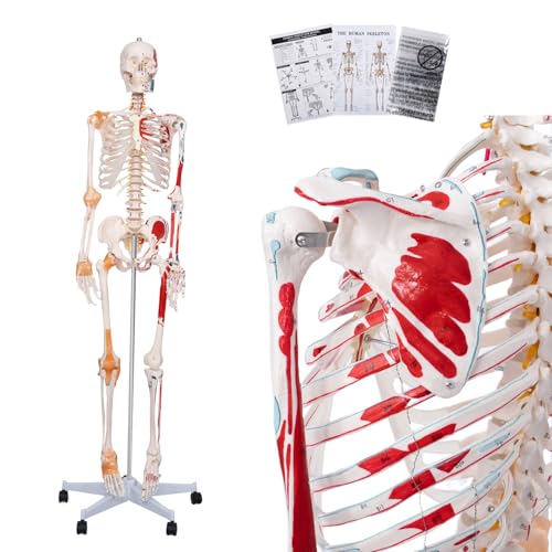
This life-size model covers the features most instructors expect from a general-purpose musculoskeletal skeleton. Painted muscle origins and insertion points provide a colorful map across the bones, while numbered structures give students something concrete to match with a poster or course notes. Flexible joint ligaments add another layer of context. During a shoulder lesson, for example, learners can look past the bone shapes and see how the joint is connected, which makes the explanation feel far less abstract.
The practical hardware is equally useful. The listing specifies hinged arms, legs, and joints, plus a removable skull cap, movable jaw, and three detachable teeth. Nervous-system and carotid-artery structures broaden its usefulness beyond a bare skeleton. A five-wheel metal stand lets an instructor roll the model beside a treatment table or between lab stations, while the included dust cover helps keep those small painted details clean during storage. That is a remarkably broad package for a single teaching model.
Pros
- Life-size 70.8-inch proportions support room-scale demonstrations
- Painted muscle origins and insertions complement numbered bones
- Flexible joint ligaments and hinged joints support movement lessons
- Removable skull cap, movable jaw, and detachable teeth allow closer study
- Five-wheel stand, dust cover, illustrations, and poster improve everyday usability
Cons
- Its full-body footprint requires more storage space than a mini or regional model
- The broad anatomical coverage is less specialized than a detailed disassemblable hand or foot model
This is the strongest all-around choice for a classroom, clinic, tutoring room, or serious home study area where one model needs to cover many lessons. Buyers focused solely on intricate foot tendons or hand neurovascular anatomy will get more concentrated detail from a regional model. For everyone else, the combination of full scale, visible attachment points, ligament representation, movable components, and storage accessories is tough to beat.
To buy this product, click here.
2. Medical Life-Size Human Skeleton Replica: Best for Medical and Nursing Instruction

Standing about 71 inches tall, this skeleton has the visual presence needed for group teaching. A student in the back row can follow a demonstration at the knee or shoulder without everyone crowding around the model. The hand-painted muscle origin and insertion points help connect skeletal landmarks to muscular anatomy, while the spinal nerves encourage broader discussions around the vertebral column. Joint ligaments supply useful context at articulations where bare bones tell only part of the story.
The listing highlights movement at the shoulder, lower jaw, elbow, wrist, knee, and foot joints. Arms and legs can be removed, and the calvarium opens for cranial study. Those features suit lab stations where one group studies an upper limb while another works through axial anatomy. The PVC surface can be wiped clean, and the detachable rolling stand has four casters. Compared with the top pick, the stand has one fewer caster and the accessory package is less extensive, but the core teaching anatomy remains compelling.
Pros
- Approximately life-size 71-inch design works well for group instruction
- Shows spinal nerves alongside muscle origin and insertion markings
- Represents joint ligaments at key articulations
- Removable calvarium, arms, and legs support focused demonstrations
- Detachable four-caster stand provides classroom mobility
Cons
- The four-caster base may feel less broadly planted than a five-wheel stand
- The listing does not specify a dust cover or the same range of study accessories as the top pick
Medical, nursing, physiology, and allied-health instructors who want a tall model with spinal-nerve detail should place this high on the list. Its articulated regions make it useful for pointing out movement pathways in front of a group, while removable limbs add flexibility at lab tables. Small apartments and private study desks are poor matches for its height, and learners seeking tiny regional structures should look to the hand or premium foot options instead.
To buy this product, click here.
3. Breesky 70.8-Inch Human Skeleton Model With Ligaments: Best for Classroom Demonstrations

For a teacher wheeling a skeleton from the anatomy lab to a neighboring classroom, stability and straightforward labeling matter every bit as much as microscopic detail. This Breesky model stands 70.8 inches tall and rides on a five-wheel base, creating a familiar full-body reference that can follow a lesson around the room. Painted muscle origins and insertions, numbered bones, flexible joint ligaments, and nervous-system structures give an instructor several visual layers to work with.
The skull is removable for a closer look at the jaw and teeth, while the arms and legs detach for regional demonstrations or easier handling. Three included posters can remain on the wall as labels are discussed on the three-dimensional model, a handy pairing for visual learners. The major caveat is the nonflexible spine. It works well for identifying vertebral regions and overall alignment, but it cannot reproduce spinal bending during a movement demonstration.
Pros
- Life-size format is easy to see during lectures and demonstrations
- Combines numbered bones with painted muscle attachment points
- Flexible joint ligaments add context around major articulations
- Detachable skull and limbs simplify focused regional teaching
- Five-wheel stand and three posters support classroom use
Cons
- The spine cannot bend, limiting demonstrations of spinal motion
- Its large rolling format is cumbersome in tight study rooms
This model makes the most sense in a classroom, clinic, or training office where it will be viewed by several people and moved regularly. The posters are useful for quick identification drills, while the detachable limbs can turn a whole-body lecture into a focused table exercise. Students who need a portable dorm-room model should choose the Axis Scientific mini skeleton, and instructors centered on detailed biomechanics may prefer the separate six-joint set.
To buy this product, click here.
4. 70.8-Inch Life-Size Human Skeleton Anatomy Model: Best for Protected Storage

Dust has a sneaky way of settling into vertebral spaces, tooth sockets, and the web of a teaching model’s ligaments. This 70.8-inch skeleton addresses that mundane but real problem with an included protective cover. It also provides the anatomical essentials expected from a feature-rich full-body model: numbered bones, painted muscle origins and insertions, flexible joint ligaments, nervous-system detail, and carotid-artery structures.
Its skull cap comes off, the jaw moves, and three teeth detach for a closer view. Hinged limbs and joints allow posing during demonstrations, while a metal stand with five rolling wheels keeps the tall structure mobile. Color illustrations, assembly instructions, and anatomy posters round out the package. The model closely resembles the top overall choice in scope, so the decision may come down to listing availability and the details a buyer values most rather than a dramatic difference in intended use.
Pros
- Includes a dust cover for cleaner long-term storage
- Painted attachment points and numbered bones aid identification
- Joint ligaments, nervous-system structures, and carotid arteries expand lesson options
- Movable jaw, removable skull cap, and detachable teeth support cranial study
- Five-wheel metal stand and hinged joints make positioning easier
Cons
- Requires substantial vertical and floor space during use and storage
- Its feature set overlaps heavily with other life-size models in this selection
Schools and clinics that leave a skeleton parked in a corner between lessons will appreciate the cover more than it first appears. Pull it off, roll the model beside a whiteboard, and the muscle markings are ready for the day’s attachment-point review. Buyers seeking maximum regional complexity should still choose a specialized hand or foot model, but this is a sensible whole-body alternative with a notably complete storage and study package.
To buy this product, click here.
5. Six-Piece Human Joint Anatomy Model Set With Ligaments: Best for Joint Mechanics

Sometimes a towering skeleton is too much model and too little interaction. This set takes the opposite approach by separating six major regions: shoulder, elbow, hip, knee, hand, and foot. Each life-size model includes connected ligaments and articulated movement, so learners can gather around a table, pick up a joint, and explore bending, rotation, or flexion from several angles. That tactile format is especially engaging during small-group biomechanics lessons.
Sturdy, lightweight PVC construction suits repeated classroom handling, and the individual pieces can be distributed across lab stations. A physiotherapy class might compare hip and shoulder mobility side by side, while a nursing student can concentrate on the knee without wrestling an entire skeleton into position. This set does not provide painted muscle origin and insertion maps or whole-body spatial relationships, so it complements a full skeleton rather than replacing one for comprehensive skeletal-system instruction.
Pros
- Includes shoulder, elbow, hip, knee, hand, and foot models
- Life-size regional designs make joint structures easier to inspect
- Articulated ligaments demonstrate bending, rotation, and flexion
- Separate pieces work well across multiple lab stations
- Lightweight PVC construction supports repeated educational handling
Cons
- Does not show whole-body alignment or relationships between distant joints
- The listing emphasizes ligaments rather than detailed painted muscle attachment maps
This is the smart pick for biomechanics courses, tabletop demonstrations, science projects, and instructors who want several students handling models at once. It is also easier to store than a standing skeleton, though six separate pieces still need an organized shelf or cabinet. Choose it for movement and articulation. Choose a life-size full skeleton when posture, axial relationships, and muscle attachment patterns across the entire body matter more.
To buy this product, click here.
6. Numbered Hand Skeleton Model With Muscles, Ligaments, Nerves, and Blood Vessels: Best for Hand Anatomy

The hand packs a dense arrangement of bones, muscles, ligaments, nerves, and vessels into a small space. On a full skeleton, those fine relationships can turn into a pale blur. This seven-piece hand model magnifies their educational importance by making the region the sole focus. Numbered structures support identification, while the modeled soft tissues help students connect the skeletal framework to the pathways crossing the palm and wrist.
Realistic textures and hand-painted assembly make the model suitable for a bright study desk, anatomy classroom, or patient-education conversation. A learner can rotate it to inspect structures from the palmar and dorsal perspectives rather than squinting at a tiny full-body hand. Its specialization is also the limitation. There is no elbow, shoulder, or whole-limb context, so anyone teaching the entire upper extremity will need another model or complementary diagrams nearby.
Pros
- Concentrates on the hand’s crowded anatomical relationships
- Shows bones, muscles, ligaments, nerves, and arteries
- Numbered structures support guided study and identification
- Seven-piece construction provides more regional detail than a basic skeletal hand
- Compact format suits desks, classrooms, and patient demonstrations
Cons
- Regional design does not show the hand’s relationship to the forearm and elbow
- Fine structures may require more careful handling than a simple one-piece bone model
Students studying hand anatomy, educators explaining common injury locations, and practitioners who need a compact visual reference are the natural audience. Its close-up detail gives it an advantage over the simplified hand attached to a life-size skeleton. Buyers building a broad introductory anatomy display should start with a full-body model, while those repeatedly returning to the palm, wrist, or digital structures will gain more from this focused format.
To buy this product, click here.
7. Foot Skeleton Model With Muscles, Ligaments, Nerves, and Blood Vessels: Best Budget Foot Model
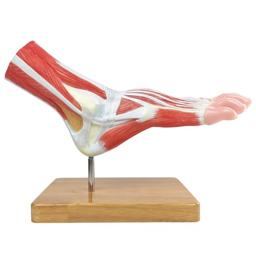
A foot skeleton becomes much more informative once the cords, bands, and colored pathways around its bones come into view. This life-size regional model represents muscles, ligaments, nerves, arteries, and skeletal anchor points, giving students a practical bridge between a bare osseous model and a crowded textbook plate. The underside is particularly valuable because the listing specifies ligament, nerve, and artery detail under the sole.
Its approachable positioning makes it attractive for students, small teaching practices, and home study spaces that cannot justify a premium disassemblable model. The form is compact enough to sit beside lecture notes without dominating the desk, yet life-size proportions keep the anatomy relatable. Compared with the 3B Scientific model below, it offers fewer documented removable components and less explicit tendon-sheath detail. That distinction matters for advanced foot and ankle coursework, but it may be unnecessary for introductory visualization.
Pros
- Life-size format provides familiar foot proportions
- Shows bones, muscles, ligaments, nerves, and arteries together
- Includes structures on the plantar surface of the foot
- Compact footprint suits personal study and small consultation rooms
- Offers a more accessible route into regional anatomy than premium specialist models
Cons
- Provides less documented disassembly than the six-part 3B Scientific model
- Does not supply whole-leg or full-body context for gait and posture lessons
This foot model fits students who want a tangible regional reference without stepping up to a highly specialized premium design. It can make a study session feel much clearer when nerves and ligaments overlap in a printed illustration. Advanced programs that need to trace individual tendons through the ankle or remove sections for deeper inspection should invest in the 3B model, while general learners may find this option comfortably sufficient.
To buy this product, click here.
8. 3B Scientific M34/1 Foot Skeleton With Ligaments and Muscles: Best Premium Regional Model

This is the model for the learner who wants to follow a tendon rather than merely see that one exists. The 3B Scientific M34/1 covers the foot and part of the lower leg, showing bones, muscles, tendons, ligaments, nerves, arteries, and veins. On the frontal side, the extensor muscles of the lower leg remain visible, and their tendons can be traced beneath the transverse and cruciate crural ligaments toward their insertion points.
Six removable parts open the model for closer study, while visible tendon sheaths add a level of regional specificity that simpler foot replicas do not document. Access to 3B Smart Anatomy courses and an available warranty extension add educational and ownership value, subject to the manufacturer’s registration terms. This depth carries a premium position and a narrow focus. It can be magnificent for a foot and ankle unit, yet it cannot replace a standing skeleton during a whole-body anatomy lecture.
Pros
- Represents muscles, tendons, ligaments, nerves, arteries, and veins
- Disassembles into six removable parts for layered study
- Shows lower-leg extensor muscles and their tendon pathways
- Includes visible tendon sheaths and detailed ankle-region structures
- Provides access to 3B Smart Anatomy learning resources
Cons
- Premium positioning may exceed the needs of introductory learners
- Regional scope does not provide whole-body skeletal relationships
Podiatry education, advanced anatomy courses, and detailed foot or ankle demonstrations are where this model shines. Picture a learner tracing a tendon with a fingertip from the lower leg, under the crural ligaments, and toward its insertion. That physical route is far easier to grasp than a stack of overlapping lines on paper. General anatomy students with a limited budget can choose the simpler foot model, but specialists will recognize the value of this richer construction.
To buy this product, click here.
9. Axis Scientific Mini Human Skeleton Model: Best Compact Option

A 31-inch skeleton can live on a desk or bookshelf where a six-foot model would become an awkward roommate. Axis Scientific preserves a whole-body view in this compact format and marks the muscle origin and insertion points with paint and numbering. That combination suits students who need to jump from a study manual to the humerus, pelvis, or femur without rolling a large stand across the room.
The shoulder, elbow, wrist, hip, knee, and ankle articulate, giving the miniature model useful movement despite its reduced scale. It comes mounted on a removable white base with a metal stand, and the included full-color manual identifies modeled features. The principal trade-off is easy to see: small anatomical landmarks are harder to inspect and demonstrate from across a classroom. The listing focuses on muscle attachments rather than modeled ligaments, so buyers requiring prominent ligament structures should select one of the life-size or regional alternatives.
Pros
- Compact 31-inch height fits desks, shelves, and smaller rooms
- Painted and numbered muscle origin and insertion points aid study
- Articulates at six major joint regions
- Removable base and metal stand provide flexible display options
- Includes a full-color identification manual
Cons
- Reduced scale makes small landmarks harder to inspect or show to a large class
- The listing emphasizes painted muscle points rather than represented joint ligaments
This is the practical choice for dorm rooms, home offices, tutoring desks, and classrooms that need a secondary reference rather than a life-size centerpiece. It gives learners whole-body orientation without a rolling base or storage cover. Large lecture rooms will expose its scale limitation quickly, and ligament-focused courses should choose a model with those structures explicitly included. For compact skeletal and muscle-attachment study, however, its balance is hard to dismiss.
To buy this product, click here.
How to Choose a Skeleton Model With Muscles and Ligaments
Start With Whole-Body or Regional Scope
A full-body skeleton shows relationships that isolated parts cannot. Students can follow the spine into the pelvis, compare upper and lower limbs, and discuss posture while the model stands upright. This is particularly useful when an instructor points from the scapula to the humerus and then down through the forearm during one continuous explanation.
Regional models trade that broad map for concentrated detail. A hand model can devote visible space to nerves and arteries that would be tiny on a standing skeleton. Likewise, a disassemblable foot and lower-leg model can show tendon pathways around the ankle with satisfying clarity. Match the scope to the lessons that will occur most often, rather than buying the largest model by default.
Check How Muscles Are Represented
The phrase “with muscles” can describe two very different products. Many skeletons use painted areas to identify muscle origins and insertions on the bones. These markings are excellent for memorizing attachment sites, but they are not three-dimensional replicas of complete muscles. A red or blue patch on the femur tells a different story than a modeled muscle belly crossing a joint.
Regional products may include raised or shaped muscle structures, tendons, and surrounding neurovascular details. Read the feature list closely and decide whether the course focuses on attachment points, soft-tissue pathways, or both. For an exam on origins and insertions, clear paint and numbering may be more useful than decorative bulk.
Look Closely at Ligaments and Articulation
Ligaments help learners understand why joint surfaces stay connected and how movement is constrained. A shoulder or knee with represented ligaments creates a much richer teaching moment than two loosely connected bones. Articulation matters too, particularly for demonstrations of flexion, extension, and rotation.
Movement claims deserve careful reading. A skeleton may have movable limbs while retaining a fixed spine. That arrangement is perfectly useful for identifying vertebrae, but it cannot demonstrate spinal flexion or lateral bending. Regional joint sets often provide better access for hands-on movement because the learner can hold each model and view it from several angles.
Consider Removable Components
Detachable limbs turn a standing skeleton into a more flexible lab tool. An instructor can place an arm on a table for a small group while keeping the axial skeleton upright for another exercise. Removable skull caps, jaws, and teeth provide similar access around the cranium, although tiny loose pieces demand organized storage.
Disassemblable regional models go further by exposing layers or pathways that are difficult to see from the surface. The six-part 3B foot model is a good example. Such construction suits advanced study, but a simpler fixed model may survive frequent beginner handling with less fuss.
Match Size to the Learning Environment
Life-size models have a wonderfully immediate presence. In a lecture room, a tall skeleton makes the angle of a hip or elbow visible from several seats away. It also occupies real floor space, needs a stable base, and may have to pass through busy hallways with arms swinging slightly as it rolls.
A mini skeleton is easier to keep beside a laptop, but smaller landmarks demand close viewing. Regional models sit between those extremes. They are life-size where it counts, yet compact enough for tabletop study. Before ordering, picture the model in its actual destination, including the base, surrounding walkways, and storage area.
Evaluate the Stand and Storage Setup
For a full-size skeleton, the stand is part of the learning experience. Four or five casters allow movement between stations, while a broad base helps the model remain upright during demonstrations. Detachable stands can simplify transport, though assembly adds another step when the model arrives.
Dust protection deserves attention in laboratories, treatment rooms, and classrooms where a model may sit untouched during school breaks. Painted markings and narrow skeletal spaces are tedious to clean one by one. A fitted or included cover is a modest accessory that can save plenty of time over years of ownership.
Use Numbering and Manuals Wisely
Numbered bones and muscle markings are most useful when paired with a clear key. Without one, a collection of tiny numbers can feel like a scavenger hunt. Posters and manuals let students identify a structure, turn back to the model, and confirm its position in three dimensions.
Color coding can speed up study, but learners should still connect each color with the correct anatomical name and relationship. The model is a visual reference, not a substitute for course materials, qualified instruction, or established anatomical resources. The strongest setup pairs physical exploration with a reliable atlas or curriculum.
Best Skeleton Models With Muscles and Ligaments FAQ
Are Painted Muscle Origins and Insertions the Same as Full Muscles?
No. Painted origins and insertions mark where muscles attach to bones, usually with color-coded areas or numbered references. Full muscle models represent more of the muscle’s three-dimensional shape and pathway. Attachment markings are especially useful for skeletal landmark study, while modeled muscles provide more context about volume and relationships with nearby structures.
Is a Life-Size Skeleton Better Than a Mini Skeleton?
Life-size models are better for room-wide demonstrations and examining structures at familiar human scale. Mini skeletons are easier to store, transport, and use at a desk. A teacher addressing a class will usually benefit from full size, while an individual studying in a bedroom or dorm may learn more comfortably from a compact model kept within arm’s reach.
Can These Models Demonstrate Realistic Joint Movement?
They can demonstrate simplified joint movement, provided the listing specifies articulation or flexible ligaments. They do not reproduce every biological constraint, tissue response, or range of human motion. Check each joint individually because a model with movable limbs may still have a fixed spine or limited movement in smaller joints.
Which Model Is Best for Studying the Foot and Ankle?
The 3B Scientific M34/1 is the most detailed foot and ankle option here. It includes six removable parts and represents muscles, tendons, ligaments, nerves, vessels, tendon sheaths, and lower-leg extensor structures. The more affordable life-size foot model is a sensible choice for introductory regional study where extensive disassembly is unnecessary.
What Is the Benefit of Detachable Arms and Legs?
Detachable limbs allow closer tabletop study and make it easier to organize separate lab activities. They may also simplify moving or storing a life-size skeleton. The trade-off is that connection points and loose components need careful handling, particularly in classrooms where many students rotate through the same station.
Are Anatomy Skeleton Models Suitable for Patient Education?
They can serve as useful visual references during general educational explanations. A clinician can point to a joint, bone, or ligament so that a patient has a clearer spatial picture. The model should remain an explanatory aid rather than a diagnostic tool, and its simplified structures cannot represent every individual anatomical variation.
How Should a PVC Skeleton Model Be Cleaned?
Follow the manufacturer’s care directions. Several models in this selection specify wipe-clean PVC, so a soft cloth is generally the starting point. Avoid aggressive scrubbing around painted markings, flexible ligaments, nerves, vessels, and tiny removable pieces. A dust cover reduces how often detailed cleaning is needed.
Do Numbered Models Always Include a Reference Key?
No. Some products include posters or manuals, while others may rely on separate course materials. Confirm what accompanies the model before purchasing. A well-matched key makes numbered bones and muscle attachment sites considerably easier to study, particularly for beginners working without an instructor nearby.
Final Verdict
Among these nine picks, the 70.8-Inch Human Anatomy Model with Muscle Insertions and Ligaments is the strongest overall option. It combines life-size proportions, painted attachment points, numbered bones, joint ligaments, nervous-system detail, movable cranial components, a rolling stand, and a dust cover. That broad toolkit makes it equally at home beside a classroom whiteboard or in a clinic where a clear visual explanation can cut through anatomical jargon.
Specialized buyers have compelling alternatives. The Axis Scientific Mini Human Skeleton is far easier to place in a small study area, while the six-piece joint set turns biomechanics lessons into hands-on tabletop sessions. For advanced foot and ankle work, the 3B Scientific M34/1 justifies its premium position with removable parts and detailed tendon pathways. Before choosing, decide whether whole-body relationships, regional detail, articulation, or compact storage matters most. The right model is the one that fits the lesson, survives the intended handling, and makes difficult spatial anatomy click when the page feels flat.
