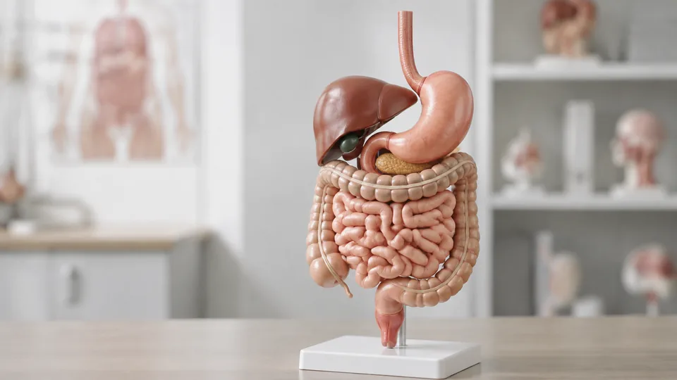
A flat diagram can make the digestive tract look like a tidy tube. Place a three-dimensional model on a desk, lift away the stomach wall, and suddenly the relationships among the liver, duodenum, pancreas, and intestines make far more sense. That jump from memorizing labels to seeing spatial anatomy is why students, teachers, and healthcare educators seek the best digestive system models.
These models differ sharply in scale, anatomical coverage, removable parts, pathology detail, and classroom durability. A compact gastrointestinal model may fit neatly beside a laptop, while a life-size display is easier for a room full of students to follow. Buyers often focus on color and overlook practical details such as a stable base, a useful identification key, or whether the structures they need are actually represented.
Different learners have different priorities. The Axis Scientific model is the strongest overall choice for comprehensive teaching, while the EISCO model offers a practical balance of detail and value. Students on a tighter budget may prefer a compact pathology model or the hands-on NewPath paper kit.
Quick Answer
Best overall: The Axis Scientific Human Digestive System Model combines broad organ coverage, removable components, hand-painted detail, and flexible tabletop or wall display.
Best value: The EISCO Hand-Painted Human Digestive System Model provides detachable parts, numbered structures, and a sturdy base without moving into the premium tier.
Best for pathology study: The Human Digestive System Model With Removable Stomach, GI Tract, and Colon emphasizes gastrointestinal and colorectal lesions in a compact format.
For a fast side-by-side view, the table below highlights the differences that matter most.
Best Digestive System Models Comparison Table
| Product | Best For | Format | Key Feature |
|---|---|---|---|
| Axis Scientific Digestive System Model | Overall teaching | Full digestive system display | Removable colon and stomach wall |
| EISCO Hand-Painted Model | Value | Two-part mounted model | Numbered structures and key card |
| EVOTECH SCIENTIFIC Model | Life-size study | Two-part life-size model | Color-coded digestive organs |
| JNMFTD Anatomy Model | Classroom demonstrations | Life-size model | Numbered anatomy and manual |
| HAMModel Digestive System Model | Premium hands-on study | Life-size multi-component model | Detachable stomach and liver |
| Removable GI Tract and Colon Model | GI pathology | Compact cross-section model | Rectal and colonic lesion detail |
| JWQFBC Digestive System Model | Small study spaces | Compact PVC model | Portable stomach and colon view |
| Menolana Pancreas Teaching Aid | Pancreas and gallbladder study | Regional pathology model | Depicts several local conditions |
| NewPath 3-D Paper Model Kit | Budget activities | Student assembly kit | Build-it-yourself learning format |
The full reviews below explain how each model fits a particular teaching environment, study level, and storage situation.
1. Axis Scientific Human Digestive System Model: Best Overall

The Axis Scientific model earns the top position through its unusually broad view of digestive anatomy. It represents the mouth, esophagus, stomach, liver, pancreas, gallbladder, intestines, and spleen, then opens the duodenum, caecum, and rectum for closer inspection. Picture an instructor tracing a bite of food from the mouth to the large intestine. The complete route remains visible without switching between several disconnected teaching aids.
Its transverse colon and front stomach wall are removable, which adds a useful hands-on layer. Students can see structures hidden behind the anterior surfaces rather than trying to imagine them from a printed cross-section. The hand-painted model sits on a base that supports tabletop and wall display, while the included full-color manual uses photographs of the model. That pairing is particularly handy during independent review, where a numbered structure means little without a clear reference close at hand.
Pros
- Covers the principal digestive organs from the mouth through the rectum
- Includes removable transverse colon and front stomach wall
- Shows open views of the duodenum, caecum, and rectum
- Can be displayed on a tabletop or wall
- Includes a full-color manual with photographs of the model
Cons
- Its comprehensive format may require more display room than a compact regional model
- Buyers studying a single condition may find a focused pathology model more direct
This is the most rounded option for an anatomy classroom, clinic education room, or serious home study station. It has enough breadth for introductory lessons and enough removable detail to support closer gastrointestinal review. A learner concerned mainly with gallstones or colon lesions should choose a specialized model, but anyone seeking one display to cover the digestive system as a connected whole will find the Axis design especially convincing.
To buy this product, click here.
2. EISCO Hand-Painted Human Digestive System Model: Best Value

For buyers who want a conventional teaching model without climbing to the highest price bracket, the EISCO strikes an appealing balance. Its hand-painted gastrointestinal tract and associated organs create clear visual separation, and detachable parts invite closer study. During a tutoring session, a student can follow a numbered component on the model, check the matching key card, and return to the physical landmark without riffling through a thick atlas.
The model is secured to a sturdy base, a quiet but valuable feature in busy classrooms. A freestanding organ piece that slides whenever someone points at it quickly becomes annoying. This mounted arrangement is better suited to demonstrations, shelf display, and repeated reference. Compared with the Axis model, its educational resources are simpler, but the numbered components and corresponding card cover the essentials cleanly.
Pros
- Hand-painted structures provide clear anatomical separation
- Detachable parts support interactive study
- Numbered components correspond with an included key card
- Sturdy mounted base suits frequent classroom display
- Balances useful detail with accessible everyday teaching
Cons
- The key card is less extensive than a full photographic manual
- Offers less specialized pathology detail than disease-focused models
EISCO is a smart fit for high school biology rooms, introductory anatomy courses, tutoring spaces, and clinics that need a dependable visual reference. Its strongest appeal lies in simplicity. The model gives learners something they can point to, detach, identify, and discuss without turning every lesson into a complicated setup. Advanced medical learners seeking deeper sectional anatomy may prefer one of the life-size alternatives.
To buy this product, click here.
3. EVOTECH SCIENTIFIC Human Digestive System Anatomy Model: Best Life-Size Option

A life-size model makes anatomical relationships easier to appreciate from several feet away, and that is where the EVOTECH SCIENTIFIC option shines. The color-coded mouth, esophagus, stomach, intestines, liver, and gallbladder create a readable pathway through the gastrointestinal system. For an instructor standing beside a lab bench, the larger format helps students connect organ position with the proportions they have seen in textbooks.
The two-piece configuration separates the digestive tract and colon for closer examination. That removable design supports a natural lesson rhythm: view the complete arrangement first, then isolate a major section for discussion. Premium PVC construction is intended to tolerate repeated educational use, making the model suitable for classrooms, clinics, and laboratories where many hands may handle it across the semester.
Pros
- Life-size presentation supports room-wide demonstrations
- Two-piece design allows the digestive tract and colon to be separated
- Color coding helps distinguish neighboring organs
- Represents major structures from the mouth through the intestines
- PVC construction is suited to recurring educational handling
Cons
- Life-size construction demands more storage and display space
- Two removable sections offer less disassembly than multi-component premium models
This model makes sense for educators who value visibility more than extreme modularity. It can anchor a lecture, support small-group lab review, or sit in a clinic room as a broad anatomical reference. Students in cramped apartments may find its scale inconvenient, while teachers addressing a group are likely to appreciate the larger, brightly differentiated structures.
To buy this product, click here.
4. JNMFTD Human Digestive System Anatomy Model: Best for Classroom Demonstrations

The JNMFTD model presents the digestive system at life size and includes the nasal cavity, pharynx, liver, stomach, rectum, and intestines. That upper-airway coverage is useful when a lesson begins before the esophagus, since learners can follow the route through the mouth and throat. The model also incorporates a coronal stomach section and transverse colon, giving the display more depth than a simple front-facing organ map.
Different anatomical areas are color coded and numbered, with a product manual supplied for identification. Imagine a practical review where one student calls out a number and another names the structure and describes its location. This model is well arranged for that back-and-forth classroom exercise. Its PVC body sits on a sturdy, detachable base and can be cleaned after repeated handling, a welcome feature in shared teaching environments.
Pros
- Life-size format is easy to present to a class
- Includes upper digestive structures such as the nasal cavity and pharynx
- Features a coronal stomach section and transverse colon
- Color-coded, numbered anatomy works with the included manual
- Detachable sturdy base supports display and storage flexibility
Cons
- Larger dimensions are less convenient for travel or desktop study
- Broad anatomical coverage may be more than young learners require
Teachers, medical educators, and students who learn through labeled visual references are the natural audience here. The combination of life-size anatomy, numbers, and a manual makes it especially useful for structured lessons and identification drills. For casual home education, a smaller and simpler model may be easier to store. For formal demonstrations, however, this model has the visual presence to hold attention across a room.
To buy this product, click here.
5. HAMModel Life-Size Human Digestive System Anatomy Model: Best Premium Hands-On Model

The HAMModel is aimed at learners who want to handle major organs and explore their spatial relationships. Its life-size display includes the esophagus, stomach, small and large intestines, liver, pancreas, gallbladder, spleen, duodenum, cecum, and rectum. It also extends upward through the nose, mouth, and throat. With that breadth, an instructor can move from swallowing to elimination while keeping the complete anatomical route in view.
Selectable parts such as the stomach and liver can be detached, making it easier to examine what sits beside or behind them. The system comprises the main digestive assembly, a coronal section of the stomach, and the transverse colon. Multiple landmarks are labeled, and the painted PVC construction arrives assembled on a stable base. There is no fiddly setup before class. Place it on the table, remove the relevant component, and the demonstration can begin.
Pros
- Life-size format presents the complete digestive route clearly
- Detachable stomach and liver support hands-on spatial study
- Includes a coronal stomach section and transverse colon
- Displays numerous upper and lower digestive structures
- Arrives assembled on a stable display base
Cons
- Premium construction represents a larger commitment than compact study models
- Its size is better suited to a dedicated classroom or lab area than a crowded desk
This is a compelling choice for medical students, biology educators, and training rooms where removable anatomy will be used regularly rather than admired from a shelf. Buyers paying for the premium format should have a clear need for life-size scale and detachable organs. A learner who mainly needs help identifying the stomach and colon can accomplish that with a smaller model, but the HAMModel offers a much richer sense of how the organs fit together.
To buy this product, click here.
6. Human Digestive System Model With Removable Stomach, GI Tract, and Colon: Best for GI Pathology

This compact model changes the focus from broad organ identification to gastrointestinal pathology. Cross-sections of the stomach, small intestine, and large intestine display common pathological morphology, while rectal and colonic lesions receive particular attention. During a patient-education conversation or pathology lesson, a raised three-dimensional feature can be easier to indicate than a tiny mark buried inside a medical illustration.
At approximately 9.8 inches tall, the PVC model sits comfortably on a desk, cart, or narrow shelf. The stomach, GI tract, and colon are removable, creating more opportunities for close viewing despite the modest footprint. An English instruction card accompanies the model. Its scale makes it easy to pass around a seminar table, though it naturally sacrifices the room-wide visibility of the larger Axis, EVOTECH, and HAMModel displays.
Pros
- Highlights gastrointestinal and colorectal pathological features
- Removable stomach, GI tract, and colon support closer inspection
- Cross-sectional presentation reveals internal morphology
- Compact height fits desks and crowded teaching carts
- Includes an English instruction card for reference
Cons
- Compact scale is less visible during large-group demonstrations
- Pathology emphasis makes it less comprehensive for whole-system introductory anatomy
Select this model for lessons centered on stomach, intestinal, rectal, or colon pathology, particularly where storage space is limited. It also suits clinics seeking a tangible communication aid, provided it is used for general education rather than diagnosis. Buyers building a foundational anatomy course should pair this type of model with a complete digestive-system reference or choose a broader option from the start.
To buy this product, click here.
7. JWQFBC Human Digestive System Model: Best Compact Option

Some learners need a model that can share desk space with a laptop, notebook, and mug of coffee. The JWQFBC measures approximately 6.30 by 4.33 by 2.17 inches, making it the most portable rigid PVC option in this group. It represents the stomach, large intestine, cecum, rectum, and duodenum, concentrating on the lower gastrointestinal area rather than the entire path from mouth to anus.
That narrow scope is part of its appeal. A tutor explaining the junction between the small and large intestines can pull out this model without hauling a life-size display from a cabinet. PVC construction supports recurring handling, and the compact body is easy to transport between a home-study space, training room, and classroom. Fine landmarks will naturally appear smaller than they do on full-size models, so viewing distance matters.
Pros
- Small dimensions suit desks, shelves, and portable teaching kits
- Represents the stomach, large intestine, cecum, rectum, and duodenum
- PVC construction supports regular educational handling
- Easy to transport between study and teaching locations
- Focused layout avoids the visual clutter of a full-body display
Cons
- Does not provide the full upper-to-lower digestive pathway
- Small structures may be difficult to see from across a classroom
This model works best for individual study, one-to-one tutoring, and demonstrations involving a few nearby learners. Its portable footprint solves a genuine problem for students without permanent lab space. Choose a life-size model for lecture halls or comprehensive organ relationships. Choose the JWQFBC when the model needs to disappear into a cabinet or travel bag after the lesson ends.
To buy this product, click here.
8. Menolana Pancreas Teaching Aid: Best for Pancreas and Gallbladder Study

The Menolana takes a regional approach, bringing the pancreas, duodenum, and gallbladder into sharp focus. Computer-coordinated coloring and hand-painted details distinguish vessels and represented lesions. Instead of scanning a sprawling digestive display for one tucked-away gland, learners can concentrate on a compact cluster of organs whose ducts and neighboring structures are closely related.
The model depicts examples of cholecystitis, gallstones, polyps, and duodenal conditions. Those visual references make it useful for pathology instruction and general educational conversations about the region. Lightweight PVC keeps the model easy to carry while resisting deformation during routine storage and handling. Since this is a specialized teaching aid, it cannot show the full progression through the esophagus, stomach, small intestine, and colon.
Pros
- Concentrates on the pancreas, duodenum, and gallbladder
- Uses contrasting colors to separate vessels and lesions
- Represents several common regional pathological conditions
- Hand-painted detail supports close visual study
- Lightweight PVC construction is easy to transport
Cons
- Regional coverage cannot replace a complete digestive-system model
- Best suited to pathology-focused or advanced lessons rather than basic organ sequencing
This is the right tool for a lesson where the pancreas and biliary structures deserve center stage. Medical educators and advanced students can use it beside a complete model to move from general orientation to close regional detail. Elementary learners or anyone studying the basic route of food through the body will gain more from a complete gastrointestinal display.
To buy this product, click here.
9. NewPath Digestive System 3-D Paper Model Making Kit: Best Budget Activity

The NewPath kit turns anatomy into a build rather than a passive display. Students assemble a three-dimensional paper model while investigating core digestive concepts, an approach that can make organ placement stick in memory. A child who has spent time cutting, folding, and positioning the stomach is far less likely to treat it as another colored blob on a worksheet.
This individual student edition includes one set of model templates and illustrated background information. The manufacturer states that the kit satisfies Next Generation Science Standards, making it especially relevant to structured school activities and home science lessons. Paper cannot match PVC for repeated handling or long-term display, but the act of construction offers a learning experience that finished models cannot replicate.
Pros
- Assembly process encourages active participation
- Includes one complete set of student model templates
- Provides illustrated background information
- Works in home and classroom learning environments
- Offers an accessible alternative to rigid anatomical models
Cons
- Paper construction is less durable than PVC
- One template set limits the kit to an individual build unless more kits are purchased
This kit is best for younger students, homeschool lessons, science projects, and teachers planning an anatomy activity rather than a permanent lab display. It rewards participation and gives the finished model a personal connection. University-level learners and educators needing precise, reusable anatomy should favor a rigid model, but few options make the basic digestive layout this approachable.
To buy this product, click here.
How to Choose a Digestive System Model
Match Anatomical Coverage to the Lesson
Start with the structures the model must teach. A complete digestive-system display should trace the pathway from the mouth and pharynx through the esophagus, stomach, small intestine, colon, and rectum. Associated organs such as the liver, gallbladder, and pancreas matter for physiology lessons, though food does not pass through them.
Regional models trade breadth for close detail. A pancreas and gallbladder model is excellent beside an advanced lesson plan, while a stomach-and-colon display keeps attention on gastrointestinal anatomy. Buying a specialized model for an introductory course can leave awkward gaps, like trying to explain swallowing while the model begins at the stomach.
Choose Between Life-Size and Compact Designs
Life-size models are easier to see from several feet away and convey realistic spatial relationships more clearly. They suit lecture rooms, labs, and clinics with dedicated display space. Their larger footprint can become a nuisance in a dorm room or shared home office, especially once books and a computer occupy the same surface.
Compact models are easier to transport and store. They work well for individual study, small tutoring groups, and mobile educators. The compromise is visibility. Tiny landmarks that look crisp at arm’s length may vanish for a student sitting in the back row.
Look for Meaningful Removable Components
Removable parts help learners see hidden surfaces and understand how organs overlap. A detachable stomach wall can expose internal anatomy, while a removable transverse colon can reveal structures positioned behind it. More pieces are not automatically better, however. Loose components can slow a short lesson and require careful storage.
Think about how the model will be used. A lecturer may prefer two large, easy-to-remove sections. A medical student reviewing organ relationships may benefit from several detachable structures that can be handled individually. Check that the removable pieces correspond to the anatomy central to your course.
Evaluate Labels, Numbers, and Reference Materials
Numbers become useful when they correspond with a clear key. Without that companion reference, a row of tiny numerals can feel like an unfinished quiz. Models with photographed manuals are especially convenient because the image orientation matches the physical display, reducing the mental shuffle between a generic diagram and the object on the table.
Printed labels can speed up basic learning, but they may clutter detailed anatomy. Numbered models often work better for formal identification exercises because learners must recall the name rather than read it directly from the organ. For independent study, prioritize a dependable key card or manual.
Consider General Anatomy Versus Pathology
General anatomy models explain where organs sit and how the digestive tract connects. Pathology models add examples of lesions, inflammation, stones, polyps, or other represented conditions. The latter can support medical education and patient communication, but they should remain educational aids rather than tools for self-diagnosis.
Beginners usually benefit from learning normal anatomy first. Once the baseline is familiar, a pathology model makes deviations easier to understand. For a clinic or advanced course, keeping one of each can create a logical progression from normal structure to condition-specific discussion.
Check the Base and Display Method
A stable base prevents the model from wobbling while someone points to a small structure. Wall-mounting capability can save counter space and keep a frequently referenced model visible. Detachable bases add storage flexibility, though instructors should keep track of mounting pieces during classroom moves.
For portable teaching, weight and shape matter as much as base stability. A broad model with projecting components can be awkward to pack despite being made from lightweight PVC. Measure the intended shelf, cabinet, or carrying case before selecting a life-size option.
Balance Durability With Learning Style
PVC models are generally better suited to repeated handling, wiping, and long-term display. Hand-painted surfaces add visual clarity but still deserve sensible care. Students should lift detachable pieces by their sturdier edges rather than tugging on narrow anatomical projections.
Paper kits occupy a different niche. They are less durable, yet assembly turns the lesson into an active project filled with folds, tabs, and small discoveries. For a one-time school activity, that tactile process may be more memorable than a premium display sitting behind glass.
Best Digestive System Models FAQ
What Is the Best Digestive System Model for a Classroom?
The Axis Scientific model is the strongest general classroom option because it covers the major digestive organs, includes removable sections, and supports tabletop or wall display. Teachers presenting to larger groups may also prefer a life-size model from EVOTECH SCIENTIFIC or JNMFTD for better visibility.
Is a Life-Size Digestive System Model Necessary?
No. Life-size models make organ relationships and demonstrations easier to see, but compact models work well for individual study and small groups. Choose based on viewing distance, storage room, and the level of anatomical detail required.
Are Removable Organs Worth Having?
Removable components are valuable when they reveal hidden surfaces or let learners explore how organs overlap. They are especially helpful for anatomy labs and hands-on learners. A fixed model can still work well for quick demonstrations where simplicity matters more than disassembly.
What Should Medical Students Look for in a Digestive Model?
Medical students should prioritize broad anatomical coverage, clear spatial relationships, internal sections, removable components, and a reliable identification manual. A general model can establish orientation, while a regional pathology model can supplement focused study of the colon, stomach, pancreas, or gallbladder.
Can Digestive System Models Be Used for Patient Education?
Yes. Three-dimensional models can help healthcare professionals point out organs and explain general anatomical relationships in accessible visual terms. They are educational aids and should not be presented as diagnostic devices or substitutes for individualized medical guidance.
What Is the Difference Between a Digestive Anatomy Model and a Pathology Model?
An anatomy model primarily represents normal structures and their positions. A pathology model depicts examples of abnormal morphology or specific conditions alongside the anatomy. Pathology models are more specialized and may omit portions of the complete digestive pathway.
How Should a PVC Anatomy Model Be Cleaned?
Follow the manufacturer’s care directions. A soft, lightly damp cloth is generally safer than abrasive pads or harsh solvents, which may affect painted surfaces. Remove detachable parts carefully, dry the model fully, and store small components where they will not be lost.
Are Paper Digestive System Models Useful?
Paper models are useful for active learning, science projects, and younger students. Building the anatomy reinforces spatial placement through repetition and physical assembly. They lack the durability and fine detail of rigid PVC models, so they are better viewed as learning activities than permanent laboratory references.
Final Verdict
Among these nine options, the Axis Scientific Human Digestive System Model is the strongest overall recommendation. Its broad organ coverage, removable transverse colon and stomach wall, open sectional views, sturdy display system, and photographic manual create a versatile package for serious study and teaching. It can carry a lesson from the mouth through the rectum without forcing the instructor to jump between unrelated visual aids.
The EISCO model is the sensible value alternative, particularly for introductory courses that need detachable, numbered anatomy on a stable base. Buyers seeking life-size, hands-on study should look closely at the HAMModel, while the compact removable GI and colon model is better suited to pathology-focused instruction. Before choosing, write down the structures your lesson must cover, measure the available display space, and decide whether removable pieces or easy portability will matter more during everyday use.
