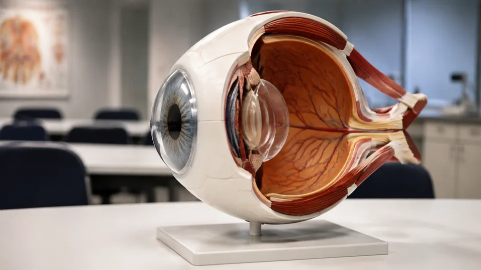
The human eye packs layers, chambers, refractive structures, and delicate tissues into a space that looks deceptively simple on a textbook page. Turn that page into a removable three-dimensional model, and the relationship between the cornea, iris, lens, vitreous body, retina, and optic nerve becomes far easier to grasp. Enlarged eye models are useful for anatomy students, science teachers, artists, healthcare educators, and anyone who learns better by lifting out a lens than by squinting at a tiny diagram.
Differences between these models run deeper than magnification. Some inexpensive 6X models focus on the basic anatomy of the eyeball, while premium orbital models add surrounding structures and finer landmarks. Removable parts, numbered structures, reference guides, stable bases, and paint detail matter more than a shiny finish. A common buying mistake is choosing the largest model without checking what separates, what is identified, or whether the stand will suit a busy classroom desk.
For a strong combination of anatomical presentation, removable construction, and supporting documentation, the Axis Scientific 5X Enlarged Human Eye Model is our top overall recommendation. Students on tighter budgets have several practical 6X alternatives, while instructors who need orbital anatomy should look toward the more elaborate EVOTECH and HADWYN models.
Short on time? The highlights below point to three especially useful options before the full comparison.
Quick Answer
- Best overall: Axis Scientific 5X Enlarged Human Eye Model, a seven-piece, hand-painted model supplied with a detailed photographic guide.
- Best labeled option: EISCO Human Eye Model, which combines seven parts with 18 numbered structures and a color key card.
- Best premium orbital model: EVOTECH Human Eye Orbital Anatomical Model, a 13-part display intended for advanced teaching and professional demonstrations.
Best Enlarged Eye Models Comparison Table
| Product | Best For | Scale | Key Feature |
|---|---|---|---|
| Axis Scientific Eye Model | Overall study and teaching | 5X | Seven pieces and photographic guide |
| Rkakneecs Eyeball Model | Budget-conscious students | 6X | Seven components and product manual |
| EISCO Human Eye Model | Numbered identification | 3X | 18 numbered structures and key card |
| EVOTECH Orbital Eye Model | Premium orbital anatomy | 3X | 13-part eye and orbit presentation |
| HADWYN Orbital Eye Model | Orbital teaching value | 3X | Numbered, handcrafted 13-part model |
| QWORK Two-Pack Eye Models | Partner and classroom activities | 6X | Two detachable seven-part models |
| faruijie Eye Model | Sagittal viewing | 6X | Seven-part median sagittal design |
| Ultrassist Eye Model | Frequent demonstrations | 6X | Detachable PVC construction and sturdy base |
| 6X Enlarged Eye Anatomy Model | Basic visual instruction | 6X | Seven removable parts on a stand |
The detailed reviews below explain where each model fits, from a quiet study corner to a lively lab table surrounded by curious students.
1. Axis Scientific 5X Enlarged Human Eye Model: Best Overall

Axis Scientific takes the top position by pairing a practical five-times-life-size format with seven removable pieces and a more polished educational package. The model separates to expose internal and external eye anatomy, allowing a student to follow the path of light from the cornea toward the lens rather than tracing that route across flat ink. Hand-painted construction gives the structures visual separation, which helps during a crowded lab session where subtle boundaries can otherwise blur together.
The full-color manual is a major strength. It uses photographs of the model, so learners can match a pictured landmark to the physical piece resting in their palm. That sounds like a small convenience until a loose lens or corneal section is sitting on the desk and the learner is trying to determine its proper orientation. Its combination of scale, dissection, and documentation suits anatomy courses, office demonstrations, and serious independent study.
Pros
- Five-times-life-size construction provides a clear view without demanding an enormous display area.
- Seven-piece design supports study of internal and external structures.
- Hand-painted details help distinguish neighboring anatomical regions.
- Full-color guide uses photographs of the actual model.
- Developed for educational use with input from medical professionals.
Cons
- Costs more than basic seven-part PVC eyeball models.
- Does not include the broader orbital presentation found on the premium 13-part picks.
This is the sensible choice for buyers who want one eye model to serve through multiple courses or teaching situations. The guide adds genuine value for self-directed learners, while the removable components make classroom explanations more tactile. Buyers focused specifically on the orbit, surrounding vessels, or a larger set of identified structures should move toward an orbital model instead. For mainstream eye anatomy, however, the Axis Scientific strikes the cleanest balance.
To buy this product, click here.
2. Rkakneecs 6X Enlarged Eyeball Model: Best Budget Pick

Students seeking a straightforward model with useful documentation should put the Rkakneecs near the top of the list. Its seven components represent the upper and lower portions, iris, cornea, lens, vitreous body, and stroma. At 6X magnification, these pieces are large enough to pass around a small study group, letting each person examine the structures before fitting them back together like a compact anatomical puzzle.
The listing specifies overall measurements of 6.2 by 6.2 inches and a 4.1-by-4.1-inch base, making this one of the easier options to carry between home and class. A detailed product manual expands its usefulness for students, teachers, artists, and anatomy enthusiasts. Picture an artist checking the shape of the iris and lens before sketching a cross-section, then packing the model into a tote. That portable, uncomplicated role is where this model makes sense.
Pros
- Six-times enlargement makes the core eye structures easy to see.
- Seven components can be removed for interactive study.
- Includes a detailed product manual for reference.
- Compact listed dimensions support storage and transport.
- Appropriate for students, teachers, artists, and general anatomy study.
Cons
- Focuses on core eyeball anatomy rather than the surrounding orbit.
- Provides less extensive identification than the EISCO model with 18 numbered structures.
Choose this model for affordable personal study, tutoring, or short tabletop demonstrations. Its manual and stated dimensions give it an edge over bare-bones models with little supporting material. Instructors leading a large lecture may prefer a more elaborate numbered model, and advanced learners may outgrow its basic scope. For carrying between a dorm room, library table, and lab bench, its compact 6X format is appealing.
To buy this product, click here.
3. EISCO Human Eye Model: Best for Labeled Study

EISCO’s model places identification front and center. Eighteen important structures are numbered on the model and correspond with an included color key card, reducing the frustrating shuffle between an unlabeled object and a separate anatomy atlas. During a review session, a study partner can call out a number while another names the structure, creating a quick drill without covering the table in flash cards.
The model is enlarged three times and divides into seven parts. EISCO describes it as a three-dimensional design developed with input from scientists, using nontoxic polymers and PVC. Hand-painted color adds a vivid, laboratory-display quality that can make borders and major regions easier to distinguish. Its 3X scale is smaller than the common 6X models, yet the numbering system and keyed reference make that compact format educationally efficient.
Pros
- Includes 18 numbered structures for systematic identification practice.
- Color key card supports quizzes and guided lessons.
- Seven-part construction allows internal anatomy to be explored.
- Hand-painted finish provides clear visual differentiation.
- Three-times enlargement balances visibility with desk-friendly display.
Cons
- Smaller than 5X and 6X models for demonstrations viewed from across a room.
- The numbering may be unnecessary for buyers seeking a simple display piece.
The EISCO is particularly well suited to students preparing for practical identification exercises and instructors who want a ready-made questioning tool. It also works nicely in a homeschool science setting, where the key card can turn a lesson into a brisk back-and-forth challenge. Buyers whose priority is maximum physical enlargement may prefer a 6X model. Those who value organized landmark identification will find this smaller model unusually purposeful.
To buy this product, click here.
4. EVOTECH Human Eye Orbital Anatomical Model: Best Premium Option

The EVOTECH steps beyond a standard split eyeball by placing the eye within a broader orbital presentation. Its 3X enlarged, 13-part design identifies structures including the cornea, sclera, choroid, retina, iris, lens, vitreous, optic nerve, fovea, ciliary body, and central retinal artery and vein. That expanded scope is helpful when a lesson shifts from basic optical anatomy toward the tissues and pathways supporting the eye.
Made from PVC, the model is described as non-toxic, odorless, durable, and easy to clean. The included stand gives it a stable presence for patient education, university instruction, or a healthcare classroom. Imagine explaining why the optic nerve exits the rear of the globe. Rather than rotating a two-dimensional diagram and hoping everyone follows, an instructor can point directly through the orbital arrangement, making the spatial relationship far more immediate.
Pros
- Thirteen-part orbital format offers broader anatomy than a basic eyeball model.
- Represents major internal structures, vessels, and the optic nerve.
- Three-times enlargement supports close tabletop teaching.
- PVC construction is described as durable and easy to clean.
- Stand supports professional display and repeated demonstrations.
Cons
- Sits in a substantially higher price tier than simple 6X eyeball models.
- Three-times scale provides less enlargement than the larger basic models.
Educators and advanced students should choose the EVOTECH when surrounding orbital context matters more than sheer magnification. Its depth would be excessive for an elementary introduction to the cornea, iris, and lens, where a seven-piece model is simpler to manage. In a university lab or professional education space, however, the extra structures can prevent a lesson from hitting the limits of a basic globe too soon.
To buy this product, click here.
5. HADWYN 3X Enlarged Human Eye Orbital Anatomical Model: Best Orbital Value

HADWYN offers another route into detailed orbital anatomy with a 13-part, three-times-enlarged format. Its represented landmarks include the cornea, sclera, choroid, retina, iris, lens, vitreous, optic nerve, fovea, ciliary body, and central retinal vessels. The wider anatomical context makes this model more informative than a simple sphere that opens to reveal a lens and vitreous section.
Different parts are marked with numbers, which is valuable during formal instruction. An educator can ask students to locate a numbered structure, then discuss where it sits relative to the retina or optic nerve. The listing also describes the model as detailed and handcrafted. Sitting on its stand, it has the visual presence expected of an advanced teaching display without relying solely on oversized dimensions to draw attention.
Pros
- Thirteen-part construction supports more advanced anatomical exploration.
- Numbered structures make guided identification easier.
- Includes orbital context absent from standard eyeball-only models.
- Represents the optic nerve, fovea, ciliary body, and retinal vessels.
- Supplied with a stand for classroom or research display.
Cons
- More complex than necessary for learners covering only introductory eye anatomy.
- Its 3X magnification is less visible at a distance than a 6X classroom model.
The HADWYN suits medical classrooms, research-oriented displays, and instructors who need to identify more than the eye’s basic refractive structures. Its numbered presentation also supports oral quizzes and structured demonstrations. A younger student may find the 13-part arrangement busy, particularly during a first lesson. For buyers comparing advanced orbital models, this is the alternative to examine when the EVOTECH exceeds the planned budget.
To buy this product, click here.
6. QWORK 6X Enlarged Human Eye Anatomy Model Two-Pack: Best for Classrooms
One model can become a bottleneck the moment several students reach for the removable lens. QWORK solves that ordinary classroom problem by supplying two 6X models, allowing partners or separate table groups to work simultaneously. Each model contains seven parts, including upper and lower eye sections, iris, cornea, lens, vitreous, and stand.
The PVC construction is described as corrosion-resistant, strong, lightweight, and easy to disassemble. Six-times magnification provides a bold view of the eye wall and principal refractive structures, which helps students connect each loose component with its position inside the globe. A teacher can disassemble one model at the front while students mirror the sequence with the second, cutting down on the awkward wait for a single teaching aid to travel around the room.
Pros
- Two-model package supports partner work and multiple learning stations.
- Six-times enlargement offers strong visibility for basic demonstrations.
- Each model consists of seven detachable parts.
- Lightweight PVC is convenient for moving between classroom tables.
- Separate stands allow both models to remain displayed at once.
Cons
- Near-identical models provide less structural variety than buying one basic and one orbital model.
- Does not offer the extensive numbering and reference key supplied with the EISCO.
This package makes the most sense for classrooms, tutoring centers, sibling study, and any setting where two learners need equal access. A single student with limited shelf space gains less from the duplicate format and may prefer the Rkakneecs model with its manual. For a science teacher setting up hands-on stations, though, two detachable models can be far more practical than one fancier display that everyone must crowd around.
To buy this product, click here.
7. faruijie 6 Times Enlarged Human Eye Anatomical Model: Best Sagittal Design
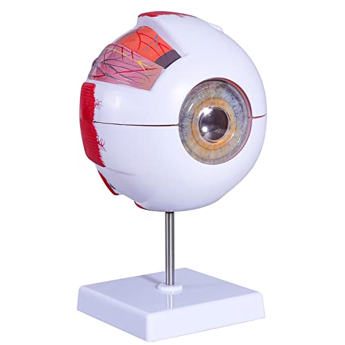
The faruijie earns its place through a median sagittal cut that presents the eye’s structures in a clean vertical section. This perspective helps learners observe the outer, middle, and inner membranes alongside the principal refractive body. It is the physical counterpart to a familiar cross-sectional textbook illustration, except the learner can pull the anatomy apart and inspect where each piece fits.
Seven removable parts comprise two eyeball sections, two vitreous sections, a lens, iris, and cornea. The listing also identifies observable muscles and part of the choroid on the sclera. Made from non-toxic PVC and paired with a stable base, it is light enough for students to handle during physiology or hygiene lessons. The crisp sectional approach is particularly useful for anyone who understands anatomy by following front-to-back alignment.
Pros
- Median sagittal section clearly exposes layered internal anatomy.
- Six-times enlargement supports easy viewing at a study table.
- Seven removable pieces encourage active assembly and review.
- Shows muscles and part of the choroid on the sclera.
- Stable base supports teaching and desktop display.
Cons
- Offers fewer identified structures than the detailed orbital models.
- Students needing a keyed labeling system will require a separate reference.
Visual learners who rely on cross-sections should find this arrangement intuitive. It is especially useful when comparing a sagittal illustration in a course guide with a tangible object. Teachers seeking numbered landmarks may find the EISCO easier for quiz sessions, while advanced anatomy courses may need orbital context. For straightforward study of the globe’s layers and refractive components, the faruijie keeps the lesson focused and hands-on.
To buy this product, click here.
8. Ultrassist Eye Anatomy Model: Best for Demonstrations
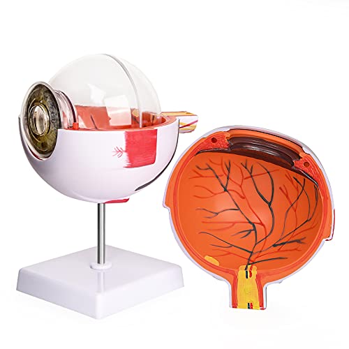
Ultrassist offers a familiar 6X eyeball format aimed at science education, medical training, and professional development. Its enlarged construction brings the internal anatomy into comfortable viewing range, whether it is sitting beneath a classroom document camera or being passed between two students. Detachable parts allow the presenter to move from the outside of the globe toward its inner structures in a logical sequence.
The model uses non-toxic PVC described as corrosion-resistant and long-lasting. A sturdy base matters during demonstrations because removing a snug component can otherwise send a lightweight display skittering across a glossy desk. Here, the stand provides a dependable home position between explanations. Its broad intended audience includes students, physiotherapists, radiology technicians, and medical practitioners using the model for educational visualization.
Pros
- Six-times enlargement produces a clear teaching display.
- Detachable construction supports sequential demonstrations.
- Sturdy base helps keep the model positioned on a desk.
- Non-toxic PVC is described as durable and corrosion-resistant.
- Suitable for science classes, study, display, and professional education.
Cons
- The available listing details do not specify an extensive numbered identification system.
- Basic eyeball scope is less suitable for teaching the surrounding orbit.
This is a practical workhorse for educators who want a stable, removable eye model without the visual complexity of a 13-part orbital display. It fits short demonstrations where the model needs to come apart, return to its base, and remain ready for the next group. Buyers wanting a photo-rich manual should favor Axis Scientific, while those planning identification drills should consider EISCO. Ultrassist is strongest as a clear, reusable presentation aid.
To buy this product, click here.
9. 6X Enlarged Eye Anatomy Model: Best Basic Visual Aid

This seven-part model covers the fundamentals without turning a first anatomy lesson into a maze of tiny labels. Its components include the upper and lower eye sections, cornea, iris, lens, vitreous, and stand. The model also presents the eye wall’s three membranes and the major refractive structures, giving students a physical way to connect the clear front of the eye with the lens and vitreous-filled interior.
Non-toxic PVC construction keeps the model lightweight, corrosion-resistant, and relatively easy to disassemble. That simplicity works well during a middle-school science unit, an introductory health course, or a quick office explanation where the goal is orientation rather than exhaustive landmark identification. Lift out the lens, point to its location behind the iris, and a concept that looked cramped in a diagram suddenly has room to breathe.
Pros
- Six-times enlargement gives basic anatomy a clear physical presence.
- Seven parts cover the principal removable eye structures.
- Lightweight PVC construction supports handling and transport.
- Stand keeps the assembled model ready for display.
- Useful for introductory science education and general anatomical study.
Cons
- Lacks the supplied photographic manual found with the Axis Scientific model.
- Less appropriate for advanced study requiring orbital structures or numerous numbered landmarks.
Buy this model for a simple, approachable visual aid that can be taken apart without introducing a long catalog of advanced anatomy. Its straightforward design suits beginners and occasional demonstrations particularly well. Serious medical coursework may warrant richer documentation or an orbital presentation, and large classrooms may benefit from the QWORK two-pack. For showing the basic geometry of the eye in three dimensions, this model stays pleasantly uncomplicated.
To buy this product, click here.
How to Choose an Enlarged Eye Model
Match the Scale to the Learning Space
A 6X model makes a strong impression on a desk and gives fingers more room to handle the lens, iris, and vitreous sections. This scale is helpful for small classroom demonstrations and learners who struggle with fine visual detail. A 3X model occupies less space and can still show substantial anatomy, especially when its landmarks are numbered or its design includes the orbit. Think about viewing distance. A student studying at arm’s length has different needs from a teacher addressing several rows of seats.
Decide Between Eyeball and Orbital Anatomy
Basic seven-part eyeball models concentrate on the globe, its layers, and major refractive structures. They are easier to assemble and less visually crowded, which makes them friendly for introductory lessons. Orbital models add a broader network of structures, potentially including the optic nerve, fovea, ciliary body, and retinal vessels. That extra detail can make an advanced lesson click, but it may leave a beginner staring at a busy cluster of colors without knowing where to start.
Check Which Components Are Removable
“Detachable” can mean different things across listings, so read the component list closely. A seven-part design commonly separates the globe, cornea, iris, lens, and vitreous sections, though exact configurations vary. Removable pieces help learners understand depth and sequence. They also introduce small parts that must be tracked, gently handled, and reassembled correctly after the lesson.
Look for Numbers, Labels, and Reference Material
A numbered model paired with a key is excellent for quizzes. Students can point, name, check, and repeat without repeatedly opening an atlas. A photographic manual is better for independent learners who need help orienting loose pieces. Models without either feature still work as visual aids, but they depend more heavily on a textbook, instructor, or separate labeled diagram for accurate identification.
Examine the Stand and Portability
A stable stand keeps the globe upright while a presenter removes components. This matters on smooth counters, where a light PVC base may shift under pressure. Portable learners should also consider the model’s dimensions, number of loose pieces, and whether the stand detaches. A compact model can travel comfortably in a tote, while an orbital display is usually happier staying on a dedicated shelf.
Balance Detail Against Clarity
More anatomy is useful only when it supports the lesson. An introductory learner may gain more from seven large, distinct pieces than from 13 closely packed structures. Medical and university teaching can justify the added complexity of an orbital model. For art reference, patient education, or general science study, clear proportions and removable core anatomy may carry more value than an extensive inventory of landmarks.
Best Enlarged Eye Models FAQ
What Scale Is Best for an Eye Anatomy Model?
For individual study and small-group teaching, 5X or 6X models offer generous visibility and easy handling. Three-times-life-size models are more compact and may include greater structural detail, numbering, or orbital anatomy. The right scale depends on viewing distance, storage space, and the complexity of the lesson.
Are Removable Eye Models Better Than Solid Models?
Removable models are generally more useful for studying internal relationships because learners can lift out the cornea, iris, lens, or vitreous components. Solid models can still serve as display references, but they provide less tactile insight into how the structures sit within the globe.
What Parts Should an Introductory Eye Model Show?
A useful introductory model should clearly represent the cornea, iris, lens, vitreous body, eye wall, and internal layers. The retina and optic nerve are also valuable landmarks where included. Beginners benefit from strong color separation and uncomplicated assembly more than from an overwhelming number of tiny structures.
Is an Orbital Eye Model Worth the Higher Cost?
An orbital model is worthwhile for advanced anatomy study, university instruction, and demonstrations involving the optic nerve, retinal vessels, ciliary body, or surrounding spatial relationships. Basic science lessons focused on light refraction and the parts of the eyeball rarely require that additional complexity.
Do Numbered Structures Make a Model Easier to Study?
Yes, especially when the model includes a matching key or manual. Numbers allow students to run identification drills and let instructors ask precise questions quickly. A model with unlabeled parts can work equally well beside a detailed course atlas, though it requires more setup during study sessions.
Can Enlarged Eye Models Be Used for Patient Education?
They can support general visual explanations in an educational setting. Large components are easier to point out than structures in a small printed image. These models remain teaching aids and do not replace individualized explanations from qualified healthcare professionals or formal medical training.
How Should an Eye Anatomy Model Be Cleaned?
Follow the manufacturer’s care directions. For many PVC models, gentle wiping with a soft, slightly damp cloth is a sensible starting point. Avoid harsh solvents, abrasive scrubbers, and soaking removable pieces unless the manufacturer specifically approves those methods, since paint and fitted connections may be affected.
Which Model Is Most Useful for a Classroom?
The QWORK two-pack is practical for parallel student activities, while the EISCO model supports structured identification through its 18 numbered landmarks and key card. For a single advanced display, the EVOTECH or HADWYN orbital model provides a broader anatomical view.
Final Verdict
The Axis Scientific 5X Enlarged Human Eye Model is the strongest all-around choice among these nine picks. Seven removable pieces, hand-painted detail, and a full-color photographic guide create a package that works for independent study as comfortably as it does for classroom explanation. It provides more educational support than a bare-bones globe without moving into the size, complexity, and premium cost of a full orbital display.
Budget-focused students should consider the Rkakneecs 6X Enlarged Eyeball Model, particularly when portability and an included manual matter. The EISCO Human Eye Model is the sharper choice for numbered identification drills, while the EVOTECH Orbital Eye Model suits advanced buyers who need broader structural context. Match the model to the lesson rather than chasing scale alone. A clear seven-piece globe can be perfect for learning the path through the cornea and lens, while orbital anatomy calls for a richer model with the landmarks to support it.
