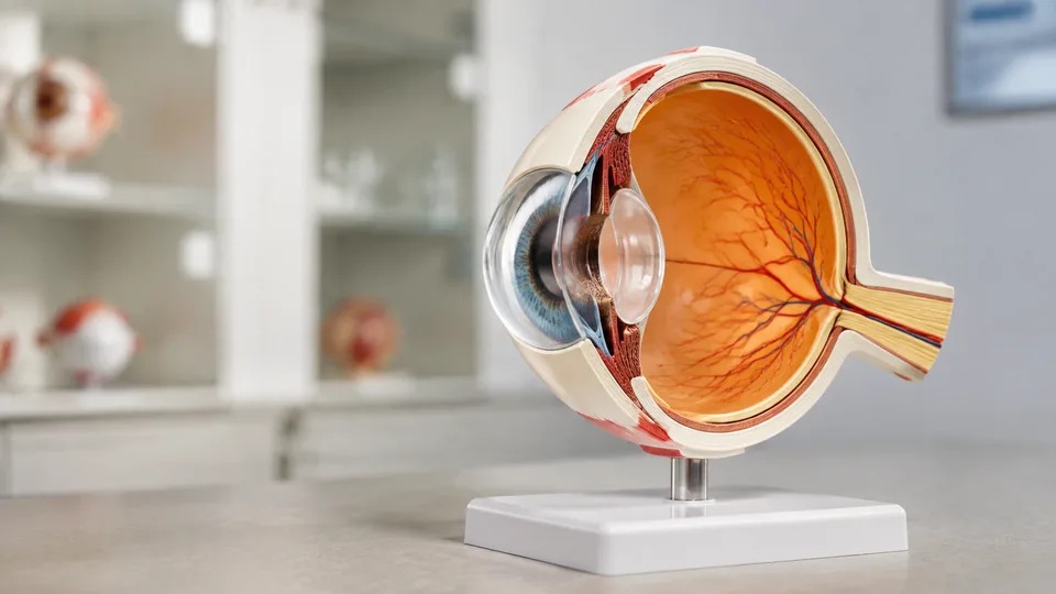
The human eye packs layers, refractive structures, delicate membranes, and an optic nerve into a space that looks deceptively simple on a printed page. Rotate an enlarged model, lift away the cornea, and suddenly the relationship among the iris, lens, vitreous body, and eye wall makes far more sense. That tactile clarity is why students, science teachers, artists, and health educators often prefer a physical eye model over another flat diagram.
Products in this category vary more than their glossy eyeball shells suggest. Some separate into seven pieces for hands-on study, while others focus on a specific condition such as cataracts. Scale, removable components, anatomical markings, base stability, and supporting guides matter most. Buying solely by appearance can leave a student with too little detail or a teacher with a model that is awkward to demonstrate across a busy classroom. For most general users, the seven-part 6X enlarged Eye Anatomy Model offers the strongest balance of visibility, straightforward disassembly, and broad educational usefulness.
For a fast look at the leading choices, the table below compares all nine models before the detailed reviews.
Quick Answer
Best overall: The 6X Enlarged Eye Anatomy Model is a versatile seven-part choice for study, teaching, and display. The GPI Anatomicals Cataract Eye Model is the standout premium option for condition-specific patient education, while the Learning Resources Cross-Section Human Eye Model is better tailored to children and introductory science lessons.
Best Eye Anatomy Models Comparison Table
| Product | Best For | Scale or Size | Key Feature |
|---|---|---|---|
| 6X Enlarged Eye Anatomy Model | Overall educational use | 6X enlarged | Seven detachable components |
| GPI Cataract Eye Model | Cataract education | 5 x 3 x 4 inches | Five interchangeable lenses |
| Learning Resources Cross-Section Eye | Children and homeschoolers | Not specified | Activity guide and labeling diagram |
| XINDAM 6X Eye Model | Guided anatomy study | 6X enlarged | Includes a labeled diagram |
| Faruijie 6 Times Eye Model | Sagittal-section study | 6X enlarged | Seven parts with visible choroid detail |
| Healthible Human Eye Model | Frequent classroom handling | 6X enlarged | Detachable ABS construction |
| Rkakneecs 6X Eye Model | Portable study | 6.2 x 6.2 inches | Detailed product manual |
| 16-Part Human Eye Model | Assembly-based learning | Not specified | Sixteen removable hand-painted parts |
| QWORK 6X Human Eye Model | Value-conscious classrooms | 6X enlarged | Reinforced display base |
The reviews below offer a closer view of how each model fits different lessons, study routines, and demonstration settings.
1. 6X Enlarged Eye Anatomy Model: Best Overall

This model earns the top position by covering the core structures most buyers expect without turning a basic eye lesson into a fiddly construction project. Its 6X enlargement makes the cornea, iris, lens, and vitreous structure easier to point out than they would be on a life-size replica. During a small-group lesson, a teacher can place it on a desk, remove the major structures, and let students trace the route of incoming light with a fingertip.
The model separates into seven components, including the upper and lower portions of the eye, cornea, iris, lens, vitreous component, and stand. The listing also identifies the three membranes of the eye wall and major refractive structures as central teaching features. Lightweight, corrosion-resistant PVC keeps it practical for repeated handling, especially where a heavy clinical display would feel excessive. Its broad usefulness is the real appeal here, stretching from introductory science study to anatomy review and patient-facing visual explanation.
Pros
- 6X enlargement gives students a clearer view of major ocular structures
- Seven detachable components support tactile, sequential learning
- Displays the membranes of the eye wall and principal refractive structures
- Lightweight PVC is easy to carry between a classroom, lab, and office
- Useful for general education, medical teaching, and anatomical display
Cons
- Does not include the condition-specific inserts found in specialized clinical models
- Seven-part construction offers less assembly depth than a model with more removable pieces
This is the sensible choice for buyers who need one adaptable model rather than a narrowly focused teaching aid. It works especially well for a college student rehearsing basic landmarks at a desk or an instructor passing components around a small lab group. Advanced ophthalmic teaching may call for finer pathological detail, but the balance of scale, separability, and approachable construction makes this the strongest general-purpose pick.
To buy this product, click here.
2. GPI Anatomicals Cataract Eye Model: Best Premium Option

General anatomy models explain where the lens sits. This specialized GPI model goes further by showing how different cataract conditions can appear within that structure. Five interchangeable lenses represent subcapsular, capsular, mature, cortical, and nuclear cataracts. Swapping those inserts during a consultation or lecture creates a crisp visual distinction that a spoken description may struggle to convey.
The lens and cornea are removable, and the model arrives with a display base plus an information card. Its eye portion measures 5 by 3 by 4 inches, while the base measures 6.5 by 5 inches, so it remains compact enough for a consultation-room shelf. The presentation feels purpose-built for healthcare education rather than broad disassembly practice. A patient can look from the clear lens to a cloudy example while an educator explains the visual concept, creating a focused, memorable demonstration.
Pros
- Five interchangeable lenses depict distinct cataract conditions
- Removable lens and cornea support focused demonstrations
- Includes an information card for convenient reference
- Compact base suits desks, consultation rooms, and display shelves
- Condition-specific design offers more relevant detail for cataract education
Cons
- Narrower teaching scope than a fully detachable general eye model
- Premium positioning may be excessive for an introductory school lesson
Clinics, healthcare educators, and instructors covering cataract types have the clearest reason to choose this model. Its interchangeable lenses provide a visual vocabulary that the general 6X models cannot match. Students seeking broad ocular anatomy should start elsewhere, since this design emphasizes lens conditions rather than taking the entire eyeball apart. For targeted patient communication, though, its specialization is precisely the point.
To buy this product, click here.
3. Learning Resources Cross-Section Human Eye Model: Best for Children

A first encounter with anatomy should invite curiosity rather than resemble a grim exam station. The Learning Resources model takes that approachable route, opening to reveal the inside of the eye and identifying its parts in a format aimed at younger learners. Picture a homeschooler opening the model at the kitchen table, matching the curved lens and colorful iris to a worksheet. That small click from picture to physical structure can make new vocabulary stick.
The included activity guide and reproducible labeling diagram give adults a ready-made way to extend the lesson. Those extras are especially useful for parents and elementary educators who want more direction than a bare plastic model provides. Its visual and tactile focus supports introductory biology, science centers, and independent exploration. This is a learning product first, so it favors accessibility and play-friendly discovery over the denser anatomical presentation expected in advanced medical study.
Pros
- Child-friendly presentation encourages hands-on biological exploration
- Opening design reveals internal eye structures
- Includes a fact-filled activity guide
- Reproducible eye diagram supports labeling exercises
- Well suited to homeschool lessons and elementary science classrooms
Cons
- Less appropriate for advanced anatomy or clinical instruction
- Product information does not specify an enlarged scale or extensive removable-part count
Parents, homeschool educators, and teachers introducing eye anatomy will get the most value from this option. The guide and reproducible worksheet reduce lesson-preparation time, while the opening eye gives children something tangible to investigate. University students who need to distinguish multiple ocular layers will want a more technical model, but younger learners are more likely to connect with this inviting cross-section than a densely detailed clinical replica.
To buy this product, click here.
4. XINDAM 6X Enlarged Human Eye Anatomical Model: Best With a Labeled Diagram

Trying to identify an unfamiliar structure on an unlabeled model can lead to that frustrating shuffle between a textbook, phone screen, and plastic eyeball. XINDAM addresses the gap by including a labeled diagram of the eye’s main parts. The reference is a modest extra, yet it matters during solo study because students can check terminology without losing their place in the physical model.
At six times life size, the model presents the major anatomy of the eyeball on a base. Its teaching focus includes the outer, middle, and inner layers of the eye wall, along with the lens and vitreous body. PVC construction keeps the model lightweight and resistant to corrosion, which is handy for educators carrying materials down a hallway. It is a straightforward anatomical study tool, but the included diagram gives it a practical advantage for learners who want labels close at hand.
Pros
- Includes a labeled diagram covering major eye structures
- 6X enlargement improves visibility during study and demonstration
- Shows the three principal layers of the eye wall
- Represents major refractive structures, including the lens and vitreous body
- Lightweight PVC construction is convenient for transport and storage
Cons
- The listing does not specify the exact number of detachable components
- Offers less condition-specific teaching value than the cataract model
This model fits anatomy students who benefit from seeing a term and its three-dimensional location together. It also makes sense for an instructor assembling a portable teaching kit, where a separate atlas would add bulk and another item to misplace. Buyers who prioritize a verified component count may prefer one of the seven-part models. For guided identification of core structures, the diagram adds welcome clarity.
To buy this product, click here.
5. Faruijie 6 Times Enlarged Human Eye Anatomical Model: Best for Sagittal-Section Study
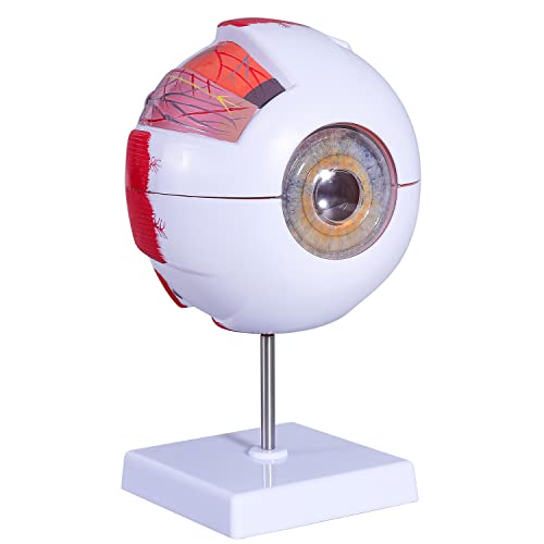
For learners trying to understand front-to-back relationships, this model’s median sagittal cut is the star feature. The section exposes the outer, middle, and inner membranes and places the principal refractive structures along a visible path. During a physiology lesson, that orientation makes it easier to follow light from the cornea through the lens toward the back of the eye without mentally flattening everything into a textbook illustration.
The eye separates into seven parts: two eyeball sections, two vitreous sections, a lens, an iris, and a cornea. The listing also identifies observable muscles and part of the choroid on the sclera, giving careful students extra landmarks to inspect. Non-toxic PVC keeps the model light, while the stable base supports desktop demonstrations. Its combination of sectioning and disassembly is especially useful for learners who like taking a structure apart, then rebuilding the anatomical sequence.
Pros
- Median sagittal cut clearly presents front-to-back anatomical relationships
- Seven removable pieces encourage active assembly and review
- 6X enlargement makes major structures easier to observe
- Shows muscles and part of the choroid on the sclera
- Stable base supports classroom and desktop presentations
Cons
- Small detachable structures require organized storage between lessons
- Does not include the labeled study diagram supplied with the XINDAM model
Visual learners studying ocular orientation should place this model high on their list. It works particularly well for demonstrating the path through the cornea, iris, lens, and vitreous region in a logical sequence. Teachers working with younger children may prefer a simpler opening model with a prepared activity guide. For secondary, college, or health-science study, its sagittal presentation makes spatial relationships refreshingly concrete.
To buy this product, click here.
6. Healthible Human Eye Model Anatomy: Best ABS Model

Many enlarged eye models use PVC, so Healthible stands apart with high-grade ABS construction. The material is described as light, sturdy, and simple to disassemble, a useful combination for a classroom prop that regularly moves from its stand into students’ hands. During a lab rotation, learners can lift out the lens or cornea, inspect the cavity, and return the pieces before the next group arrives.
The 6X model allows removal of the upper and lower eye sections, cornea, iris, lens, and vitreous component. A detachable display base provides a practical home between demonstrations and makes the model easier to present on a desk. Its major ocular structures are intended for anatomy classes, university study, and patient education. The overall concept remains familiar, but buyers who prefer ABS over the more common PVC alternatives have a clear reason to favor it.
Pros
- High-grade ABS construction offers an alternative to PVC models
- 6X enlargement supports clear viewing of major eye anatomy
- Multiple removable structures allow deeper internal inspection
- Detachable base simplifies setup and transport
- Suitable for classroom, university, and educational consultation settings
Cons
- No included labeling sheet or activity guide is specified
- General anatomy focus does not depict distinct eye conditions
Healthible is a practical fit for buyers who expect students to handle the model frequently and specifically want ABS construction. Its removable structures cover the familiar sequence needed for basic anatomy demonstrations, and the detachable base makes packing less awkward. Learners who rely heavily on printed labels should pair it with a separate diagram. Within a busy teaching environment, its simple setup and sturdy material profile are the chief attractions.
To buy this product, click here.
7. Rkakneecs 6X Enlarged Eyeball Model: Best Portable Option

A model that lives in a tote bag rather than a permanent lab needs manageable dimensions and simple disassembly. The Rkakneecs model measures 6.2 by 6.2 inches, paired with a 4.1 by 4.1-inch base, so it can move between a study desk, classroom, and presentation table without demanding much space. The included product manual offers additional guidance once the pieces come out of the package.
Its seven components cover the upper and lower portions, iris, cornea, lens, vitreous body, and stroma. At 6X magnification, the model is large enough to expose the principal structures while remaining relatively compact. The listing positions it for students, teachers, doctors, artists, and anatomy enthusiasts, which reflects its balanced design. An art student studying the eye’s rounded volumes may value the same removable lens that a science student uses to review refraction.
Pros
- Specified 6.2 by 6.2-inch model size is manageable for travel and storage
- Seven components provide useful hands-on anatomy study
- 6X magnification keeps major structures visible
- Includes a detailed product manual
- Compact base occupies limited desk or shelf space
Cons
- Compact proportions may be less visible during room-wide demonstrations
- Small loose components need careful packing during transport
Tutors, traveling educators, artists, and students with limited shelf space are the natural audience for this model. Its known dimensions make planning storage easier than with listings that omit size information. A lecturer addressing a large room may prefer a bigger, more conspicuous display. For close study at a table, though, the combination of seven components, a manual, and a modest footprint is highly practical.
To buy this product, click here.
8. 16-Part Human Eye Anatomical Model: Best for Assembly-Based Learning

Taking a model apart can turn passive observation into a spatial puzzle, and this option leans hard into that experience. The listing specifies 16 removable, hand-painted parts with an assembly guide and display stand. Instead of lifting away a handful of large sections, learners can spend more time understanding how numerous pieces relate, then work through the satisfying process of fitting the eye back together.
The plastic model is described as lightweight, detachable, and easy to clean. Those qualities suit supervised classroom projects, home study, and art-reference sessions where the model may receive frequent handling. Its higher component count is the clear differentiator among these nine picks. A student who remembers structures by building and reconstructing them may find this format more engaging than staring at a completed display, especially during a long review session.
Pros
- Sixteen removable parts provide the highest stated component count here
- Hand-painted pieces support visual separation of structures
- Assembly guide helps users reconstruct the model
- Display stand keeps the completed eye organized for reference
- Easy-to-clean plastic suits repeated educational handling
Cons
- More pieces create greater potential for parts to be misplaced
- The listing does not provide a clear scale or assembled dimensions
This is the most compelling choice for learners who absorb anatomy by assembling it piece by piece. It can transform a quiet study session into an active reconstruction exercise, though the extra components demand a tray, container, or consistently tidy storage routine. Teachers seeking a quick two-minute demonstration may find seven-part models easier to manage. For patient, hands-on learners, the 16-part format offers a deeper building experience.
To buy this product, click here.
9. QWORK 6X Human Eye Anatomy Model: Best Value

QWORK keeps the formula straightforward: enlarge the eye six times, separate it into seven sections, and give it a reinforced base for steady desktop display. That stable footing is useful during a lively science lesson, where elbows bump tables and models get turned repeatedly toward different students. The cornea, iris, lens, vitreous body, and basic anatomical layers remain clearly represented for foundational instruction.
PVC construction makes the model light enough to move around, and the detachable design supports both observation and assembly practice. It is aimed at science classes, lab study, and educational exhibits rather than condition-specific instruction. Buyers seeking an accessible general model may appreciate its lack of fuss. It covers the essential landmarks, permits internal exploration, and sits securely when the lesson shifts back to the whiteboard.
Pros
- 6X scale gives learners a clear view of core anatomical structures
- Seven detachable sections support interactive study
- Reinforced base helps keep the model stable during demonstrations
- PVC construction is lightweight for classroom transport
- Well matched to introductory lessons, labs, and educational displays
Cons
- No printed manual, labeling diagram, or activity guide is specified
- Basic structure set is less specialized than pathology-focused alternatives
Budget-conscious teachers and students who need a conventional enlarged eye model should consider QWORK. It offers the scale and removable construction that matter most for basic lessons, with a reinforced base that feels especially relevant in shared spaces. Buyers expecting extensive labels or pathological inserts should look toward XINDAM or GPI. For uncomplicated classroom visualization, this model delivers the right fundamentals.
To buy this product, click here.
How to Choose an Eye Anatomy Model
Match the Model to the Lesson
Start with the lesson rather than the shiniest eyeball on the shelf. General anatomy study calls for removable representations of the cornea, iris, lens, vitreous body, and eye wall. A healthcare educator explaining cataracts has a different task and will gain more from interchangeable pathological lenses. For a child meeting ocular anatomy for the first time, an activity guide and simple opening cross-section can be more useful than sixteen loose parts scattered across the kitchen table.
Scale and Viewing Distance
Six-times enlargement is common among the models in this guide because it makes compact structures easier to see and manipulate. That scale works well for desk study and small teaching groups. Room size still matters. A model that looks bold from two feet away may become a colored dot at the back of a lecture hall. Check specified dimensions where available, then picture the actual viewing distance in your classroom, office, or studio.
Removable Components
Seven-part models strike a useful balance between anatomical separation and manageable cleanup. They usually let learners remove the cornea, iris, lens, vitreous structure, and larger eye sections without creating a jigsaw puzzle. Higher component counts reward patient learners who enjoy reconstruction, but every extra piece needs storage. A shallow tray can prevent a tiny lens from rolling under a cabinet with that dreaded little plastic clatter.
Labels, Manuals, and Activity Materials
An unlabeled physical model works best beside a textbook or anatomical chart. Included diagrams and manuals reduce that dependency, especially for independent learners. Children benefit from guided activities and reproducible worksheets, while college students may prefer a concise labeled reference beside the stand. Built-in printed labels were not specified for most options here, so inspect the included learning materials before assuming the model can teach terminology by itself.
Material and Cleaning
PVC is common because it is lightweight, strong, and resistant to corrosion. ABS provides another light, sturdy option. For a personal study model, either material may serve comfortably. Shared classroom pieces face marker-stained fingers, dust, and frequent disassembly, making wipe-clean surfaces more valuable. Follow the seller’s care guidance and avoid harsh cleaners that could dull painted details or loosen markings.
Base Stability and Portability
A wide or reinforced base keeps the eye steady while an instructor points to the iris or removes a section. Detachable stands are convenient for storage, although separate pieces create one more item to track. Traveling tutors and students should favor known compact dimensions, lightweight construction, and straightforward assembly. Permanent displays can prioritize a polished base and clear sight lines over bag-friendly proportions.
General Anatomy Versus Pathology
Standard models explain normal structure and spatial relationships. Pathology models focus on a condition, often sacrificing broad disassembly for more targeted visual examples. Neither format replaces formal instruction or clinical expertise. Think of the model as a three-dimensional reference that supports a lesson. A general eye belongs in an anatomy lab, while interchangeable cataract lenses make more sense beside a focused educational conversation.
Best Eye Anatomy Models FAQ
What Scale Is Best for an Eye Anatomy Model?
A 6X enlarged model is a practical choice for desk study, small-group teaching, and hands-on demonstrations. It makes structures such as the lens and iris easier to handle while remaining compact enough for a shelf. Large-room teaching may require a bigger display or a projected image alongside the physical model.
Which Parts Should Be Removable?
For general study, look for removable eye halves, cornea, iris, lens, and vitreous component. These pieces help learners see the order of structures from the front of the eye toward the retina. More advanced assembly models may separate additional structures, but extra pieces require more careful organization.
Are Eye Anatomy Models Suitable for Children?
Yes, provided the model is age-appropriate and used with adult supervision where small parts are present. A simple cross-section with an activity guide is easier for younger students to understand. Complex multi-part models are better suited to older learners who can handle and store the pieces responsibly.
Do Eye Models Usually Come Labeled?
Some include a labeled diagram, product manual, information card, or worksheet rather than printing labels directly on the model. Check the listed educational materials before purchasing. Independent students often benefit from keeping a labeled reference beside an unlabeled model.
Is a Pathological Eye Model Better Than a General Model?
That depends on the teaching goal. A general model is more useful for learning normal eye structure and spatial relationships. A pathological model is stronger for explaining a particular condition, such as cataracts. Most first-time anatomy students should begin with a general detachable model.
How Many Removable Parts Are Enough?
Seven parts are enough for many introductory and intermediate lessons because they expose the eye’s principal internal structures without making assembly cumbersome. A 16-part model offers a more involved reconstruction exercise. The higher count is useful only when the additional handling supports the learner’s goals.
Can an Eye Anatomy Model Be Used for Drawing Reference?
Yes. Artists can use an enlarged eye model to study spherical form, the placement of the iris and cornea, and the relationship between visible and internal structures. Models showing extraocular muscle markings may add context, though a full head or orbital model would provide more information about how the eye sits within surrounding anatomy.
How Should Removable Eye Model Parts Be Stored?
Keep all components together in a lidded container, divided organizer, or labeled bag near the stand. A shallow tray is useful during lessons because rounded pieces can roll. Reassemble the model after study when practical, then check that the lens, iris, and cornea are present before putting it away.
Final Verdict
The 6X Enlarged Eye Anatomy Model is the strongest overall choice for most buyers. Its seven detachable components, enlarged scale, lightweight PVC construction, and coverage of major ocular structures make it useful across classrooms, study desks, and educational consultation settings. It offers enough physical interaction to clarify spatial anatomy without burdening users with a sprawling collection of tiny pieces.
Different priorities point toward worthy alternatives among these nine models. The GPI Anatomicals Cataract Eye Model is the premium specialist for cataract education, while the Learning Resources Cross-Section Human Eye Model gives children a friendlier entry point. Buyers seeking value can turn to the stable, seven-section QWORK model. Match the level of detail, part count, supporting materials, and viewing distance to the actual lesson, then choose the model that makes those tricky ocular relationships click into place.
