
The urinary system looks simple on a textbook page, yet its spatial relationships can become slippery the moment kidneys, ureters, vessels, bladder, and nearby reproductive organs pile into one diagram. A physical model turns those overlapping lines into shapes that students can rotate, point to, and remember. The differences between models are substantial, ranging from focused urinary displays to life-size pelvic cross-sections and removable, multipart teaching tools.
Buyers often focus on vivid colors while overlooking the features that influence daily use. Scale matters during a lecture, removable parts help with close study, and numbered structures paired with a clear manual can save hours of squinting at unlabeled anatomy. A nursing student reviewing at a desk has different priorities from an instructor presenting across a busy lab, while a urology office may need a model that makes patient explanations feel less abstract.
Among these nine options, the Axis Scientific Male Urinary System Model offers the strongest overall balance. Its life-size presentation, removable bladder, 19 numbered structures, photo-based manual, and three-year warranty give it unusually broad appeal for structured study and teaching.
Quick Answer
Best overall: The Axis Scientific Male Urinary System Model combines life-size anatomy with numbered landmarks and a removable bladder. The SFXYJ Male Urinary System Anatomy Model is a sensible value-oriented alternative for general demonstrations, while the PacuM Teaching Model is the premium classroom pick for buyers who want a six-part slab-style display.
For a fast side-by-side view, the table below highlights the scale, removable features, and teaching focus of each selection.
Best Urinary System Models Comparison Table
| Product | Best For | Format | Key Feature |
|---|---|---|---|
| Axis Scientific Male Urinary System Model | Overall study and teaching | Life-size urinary model | 19 numbered structures and removable bladder |
| SFXYJ Male Urinary System Anatomy Model | Value-focused buyers | Life-size model on PVC base | Removable organs and included manual |
| Female Reproduction and Urinary System Model | Female urogenital anatomy | Life-size, three-part model | Urinary and reproductive structures together |
| EVOTECH SCIENTIFIC Male Urogenital Model | Male urogenital anatomy | Life-size, three-part model | Removable construction and vascular detail |
| Human Urinary System Anatomy Model | Hands-on organ study | 1:1-scale detachable model | Removable kidneys, adrenals, ureters, and bladder |
| Merinden Male Pelvis Anatomy Model | Male pelvic relationships | Life-size, five-part half-pelvis | Magnetic parts and wall-mountable baseboard |
| Merinden Female Pelvis Anatomy Model | Female pelvic relationships | Life-size, two-part half-pelvis | Removable rectum and numbered anatomy |
| PacuM Urinary System Teaching Model | Premium classroom display | Six-part slab-style model | Numbered indicators and hand-painted structures |
| Simedutech Prostate Anatomy Model | Prostate pathology lessons | Detachable pathology model | Normal, enlarged, and cancer-related comparisons |
The detailed reviews below explain where each model fits, from quiet desk study to lively classroom demonstrations.
1. Axis Scientific Male Urinary System Model: Best Overall

Numbers and labels can make the difference between a colorful desk ornament and a serious study aid. This Axis Scientific model identifies 19 structures spanning the adrenal glands, kidneys, ureters, bladder, and urethra. Those numbers correspond to a full-color manual that uses photographs of the actual model, so students are not forced to translate a stylized drawing while holding a completely different object.
The removable bladder gives the model its strongest practical advantage. Detaching it exposes internal landmarks that include the trigone, ureteral openings, and internal urethral orifice. Picture a student preparing for a lab practical late at night. Instead of mentally peeling away layers from a flat illustration, the student can lift the bladder and trace where the structures sit. Life-size proportions also make it useful during patient education, where realistic scale can keep an explanation grounded and clear.
Pros
- Displays 19 numbered structures across the male urinary system
- Life-size proportions support realistic scale and spatial reference
- Removable bladder reveals several important internal landmarks
- Full-color manual uses photographs of the model itself
- Includes a three-year warranty and access to US-based support
Cons
- Focuses on male urinary anatomy rather than offering interchangeable male and female anatomy
- Only the bladder is specified as removable, so it is less modular than multipart organ models
This is the strongest choice for students, instructors, and clinics seeking a balanced model rather than a highly specialized pelvic cross-section. The numbered guide adds structure to independent review, while the detachable bladder creates a satisfying “aha” moment during lessons about internal anatomy. Buyers needing reproductive anatomy or multiple removable organs should look farther down the list, but general urinary-system teaching is where this model earns its top position.
To buy this product, click here.
2. SFXYJ Male Urinary System Anatomy Model: Best Value

For buyers trying to stretch a departmental supply budget, the SFXYJ model covers the essential urinary pathway without drifting into a complicated full-torso display. It depicts the kidneys, adrenal glands, ureters, bladder, arteries, and veins at life size. Distinct colors and clear textures separate neighboring structures, which helps a lecturer point across a table without every vessel blending into a reddish tangle.
The kidneys, ureters, bladder, and adrenal glands are described as removable, opening more possibilities for hands-on review than a fixed display permits. A small study group could pass an organ around, inspect its shape, and then rebuild the arrangement on the sturdy PVC base. The included product manual supports identification, while the easy-clean PVC construction suits classrooms where fingerprints, dry marker dust, and constant handling are part of the scenery.
Pros
- Represents the main male urinary organs at life size
- Kidneys, adrenal glands, ureters, and bladder are removable
- Color differentiation makes major structures easier to follow
- Includes a manual for guided teaching and study
- Sturdy base helps keep the display stable during demonstrations
Cons
- The listing does not specify the same numbered landmark count as the Axis Scientific model
- Broad external anatomy offers less pathology detail than a specialized prostate model
This model suits medical and nursing students, tutors, health-science classrooms, and clinicians who want a straightforward visual reference. Its removable components are especially useful for learners who remember shapes through touch and assembly. The Axis model has a more explicitly documented labeling system, but SFXYJ provides a compelling mix of life-size scale, modular organs, and basic guidance for value-conscious buyers.
To buy this product, click here.
3. Human Female Reproduction and Urinary System Medical Anatomical Model: Best Female Urogenital Model
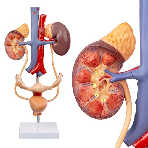
Female urinary anatomy becomes easier to understand once it is viewed alongside the reproductive system rather than floating alone on a blank board. This life-size model brings the kidneys, adrenal glands, ureters, bladder, major vessels, and female reproductive structures into one visual field. That broader context is useful for anatomy courses where students must grasp proximity and orientation, rather than memorize isolated organ names.
Three removable sections create a more active lesson. An instructor can disassemble the model during a demonstration, pause while students identify a structure, and then reconstruct the anatomy with the class. Clear textures and vascular coloring add visual separation, while the stable base keeps the arrangement upright during presentations. The included manual supplies labeled diagrams, which can help beginners move from the bold, glossy shapes of the model back to formal anatomical terminology.
Pros
- Combines female urinary and reproductive anatomy in one display
- Life-size format provides a realistic reference for organ scale
- Three-part construction supports disassembly and close inspection
- Shows kidneys, adrenal glands, ureters, bladder, arteries, and veins
- Includes a labeled instruction manual and stable base
Cons
- The combined urogenital focus may be broader than buyers seeking a urinary-only model require
- Three removable sections provide less segmentation than larger multipart teaching models
This is a natural fit for educators covering female urogenital anatomy, students comparing male and female structures, and clinics seeking a visual communication tool. Someone studying the renal system alone may prefer a model with numbered kidney and bladder landmarks. For lessons where anatomical context matters, however, seeing the urinary and reproductive structures share the same compact space is the central advantage.
To buy this product, click here.
4. EVOTECH SCIENTIFIC Male Reproduction and Urinary System Model: Best Male Urogenital Model

Male urinary structures do not exist in a neat vacuum, and this EVOTECH SCIENTIFIC model embraces that reality. Its life-size presentation includes the urinary and reproductive systems, with visible kidneys, adrenal glands, ureters, bladder, arteries, and veins. During a lecture, that combined arrangement can clarify relationships that become muddy when students flip between separate textbook plates.
The model separates into three parts for closer inspection. That modest degree of modularity keeps setup manageable while still allowing an instructor or learner to lift sections away from the display. PVC construction makes it light enough for routine movement between a cabinet and teaching bench, and the sturdy base provides a stable platform once the lesson begins. A detailed manual with labeled diagrams adds welcome direction for self-study sessions where an instructor is not nearby.
Pros
- Integrates male reproductive and urinary anatomy
- Life-size design helps communicate realistic organ proportions
- Three removable parts encourage interactive demonstrations
- Vascular structures and major urinary organs are visibly represented
- Includes a labeled manual and sturdy display base
Cons
- Students focused solely on urinary anatomy may find the reproductive detail distracting
- The listing does not specify individually numbered structures on the model
Choose this model for male urogenital courses, patient-friendly demonstrations, or comparative anatomy lessons that pair it with a female model. It is more context-rich than a urinary-only display and easier to handle than a large torso model. Learners preparing for identification-heavy practical exams may prefer the Axis model’s documented numbering, while visual learners studying organ relationships should appreciate EVOTECH’s wider anatomical scene.
To buy this product, click here.
5. Human Urinary System Anatomy Model, 1:1 Life Size: Best for Removable Organ Study

Some learners need to pull anatomy apart before the connections click. This 1:1-scale male urinary model allows the kidneys, adrenal glands, bladder, and ureters to be disassembled and reassembled. The result is especially useful for exploring the independent form of each organ, followed by the positional puzzle of putting the complete urinary pathway back together.
The model uses PVC that the listing describes as easy to clean and reusable, practical qualities in a lab where objects are handled by one group after another. Clear contours and a realistic feel support close desk study, while the complete arrangement can remain on display in a classroom, study room, or medical office. For an instructor explaining how a ureter links kidney to bladder, removing and replacing that pathway gives the lesson more physical clarity than pointing at a printed line.
Pros
- Uses a 1:1 adult male urinary-system scale
- Core organs can be removed and reassembled
- Supports study of individual organ shape and overall positioning
- Easy-clean PVC construction suits repeated educational handling
- Useful for classrooms, student study, and visual patient explanations
Cons
- The listing does not specify an included numbered reference manual
- Urinary focus provides less pelvic context than the Merinden cross-section models
This option works best for tactile learners and instructors who want students to rebuild the urinary arrangement during a lesson. Its appeal lies in organ removal rather than dense labeling or pathology comparison. Buyers wanting guided self-study should confirm whether the supplied documentation meets their needs, while those comfortable using a separate anatomy atlas can make good use of the clean, modular layout.
To buy this product, click here.
6. Merinden Male Pelvis Reproductive Anatomy Model: Best for Male Pelvic Relationships

A full urinary display is excellent for tracing urine from kidneys to bladder, but it cannot always explain what happens inside the crowded pelvis. Merinden approaches the subject through a life-size half-pelvis showing the bladder, reproductive organs, removable genital structures, and muscular anatomy. The cutaway view feels more like opening a textbook illustration into three dimensions, with depth and neighboring tissues suddenly visible.
Five parts and magnetic connections make assembly feel tidier than loose-fitting pieces. During a small seminar, the model can sit on its baseboard while an instructor removes a component and discusses the space behind it. The board may also be mounted on a wall, a handy option in a compact classroom where counter space disappears quickly. A colorful educational booklet identifies the anatomical features represented on the model.
Pros
- Shows urinary, reproductive, and muscular anatomy in pelvic context
- Life-size half-pelvis format reveals spatial relationships clearly
- Five-part construction includes removable genital structures
- Magnetic connections simplify removal and reassembly
- Baseboard supports tabletop or wall-mounted display
Cons
- Does not provide the complete kidney-to-bladder overview of a full urinary-system model
- Its sectional format is more specialized than a general introductory display
This Merinden model belongs in pelvic anatomy lessons, reproductive-system courses, and offices where the relationship between bladder and surrounding male structures needs a clear visual explanation. It should complement rather than replace a complete urinary model for renal study. Students concentrating on the lower urinary tract, however, may gain more from this revealing side view than from another front-facing organ display.
To buy this product, click here.
7. Merinden Female Pelvis Reproductive Anatomy Model: Best Female Pelvis Cross-Section

Female pelvic anatomy can look impossibly cramped in a small diagram. This life-size Merinden half-pelvis gives the bladder, reproductive organs, rectum, and muscular structures enough visual space to make their positions legible. The anatomy is numbered and painted in distinct colors, so the viewer can follow boundaries without losing the bladder among adjacent tissues.
The model separates into two parts and includes a removable rectum, exposing areas that would otherwise remain hidden in the sectional view. Magnetic assembly keeps the interaction simple, and the baseboard can stand on a teaching surface or be mounted to a wall. Imagine a student comparing pelvic compartments before an exam. The model’s physical depth makes anterior and posterior relationships far more tangible than a page covered in arrows.
Pros
- Displays female urinary, reproductive, and muscular anatomy together
- Life-size half-pelvis format clarifies crowded pelvic relationships
- Numbered and color-painted structures support identification
- Removable rectum provides a clearer view of surrounding anatomy
- Magnetic parts and mountable baseboard simplify classroom display
Cons
- Two-part construction offers fewer removable sections than the male Merinden model
- Kidneys and the complete upper urinary pathway are outside this pelvic model’s focus
Female pelvic anatomy instructors, students, and clinics are the clearest audience for this model. It offers more sectional context than the three-part female urogenital model, while that model gives a more complete view of the kidneys and ureters. The deciding question is simple: do you need the whole urinary pathway, or a sharper look at how the bladder sits within the female pelvis?
To buy this product, click here.
8. PacuM Urinary System Teaching Model: Best Premium Classroom Display
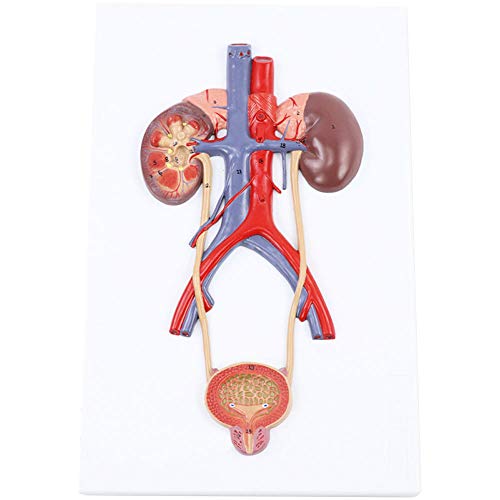
The PacuM model is aimed at buyers who want a prominent teaching display rather than a basic study prop. Its slab-style design presents the major urinary structures together, including the kidneys, ureters, bladder, arteries, and veins. Numbered indicator markings help an instructor direct attention to specific areas during a lecture, especially when a dozen pairs of eyes are scanning the same busy anatomy.
Six-part construction adds interactive value, while computer-assisted color matching and hand-painted finishing give the organs clear visual separation. In a urology classroom, the display could remain upright at the front of the room and then move onto a table for closer group discussion. PVC construction supports repeated educational use, though this model’s premium positioning makes the most sense for departments and professional teaching spaces that will use it regularly.
Pros
- Six-part design supports more detailed classroom demonstrations
- Numbered indicators make guided identification more efficient
- Shows kidneys, ureters, bladder, arteries, and veins
- Slab-style layout keeps the full urinary pathway visible together
- Hand-painted, color-separated structures create a strong display presence
Cons
- Premium positioning may exceed the needs of an individual student seeking a simple desk model
- The listing provides limited detail about the accompanying reference materials
This model is best suited to an anatomy lab, urology classroom, training room, or professional office where visibility and repeated demonstrations matter. Independent learners can find more guided documentation in the Axis Scientific option, while instructors drawn to a larger visual centerpiece may prefer PacuM’s six-part, numbered presentation. Its value depends heavily on frequent use, so occasional learners should weigh that trade-off carefully.
To buy this product, click here.
9. Simedutech Prostate Anatomy Model: Best for Pathology Comparison

Most models in this roundup teach normal gross anatomy. Simedutech takes a narrower, pathology-oriented route by comparing a normal prostate with representations of benign prostatic hyperplasia and prostate cancer. The model also displays the bladder neck, urethra, and surrounding tissues, helping students visualize why changes in the prostate can matter to the lower urinary pathway.
The detachable design invites side-by-side inspection rather than passive viewing. A teacher can place the normal and altered structures in sequence, allowing learners to compare form without flipping between separate images. Hand-painted PVC construction supplies color and texture, while the compact subject matter makes the model convenient for a desk, consultation space, or focused pathology station in a lab.
Pros
- Compares normal prostate anatomy with common pathological changes
- Includes representations of benign enlargement and prostate cancer
- Shows the bladder neck, urethra, and nearby tissues
- Detachable construction supports close comparative study
- Durable hand-painted PVC suits repeated educational handling
Cons
- Covers a specialized portion of the male urinary and reproductive systems rather than the complete urinary tract
- Less appropriate as a first model for learning kidneys, ureters, and overall urinary anatomy
This focused model makes sense for pathology lessons, urology education, and demonstrations centered on the prostate’s relationship to the urethra and bladder neck. It should be treated as a specialized supplement rather than a complete urinary-system reference. Buyers establishing a basic anatomy collection should begin with a full urinary model, while instructors adding pathology comparison will find this pick meaningfully different from the other eight.
To buy this product, click here.
How to Choose a Urinary System Model
Match the Anatomical Scope to the Lesson
Start with the structures that must be visible. A complete urinary model should generally show the kidneys, ureters, bladder, and relevant vessels, while some models also include adrenal glands or the urethra. That broad view works well for introductory anatomy and physiology. Pelvic cross-sections sacrifice the upper urinary pathway to reveal the tight relationship between the bladder, reproductive organs, rectum, and musculature.
Think about the moment the model will be used. A student tracing urine formation and transport needs the complete kidney-to-bladder route. An instructor explaining lower urinary anatomy may gain more from a male or female half-pelvis, where the crowded arrangement becomes visible at a glance.
Choose an Appropriate Scale
Life-size models offer intuitive scale, making them useful for medical education and visual explanations. They also fit more easily on a desk or laboratory bench than enlarged displays. Across a large classroom, however, realistic size can look surprisingly small, especially around narrow ureters and vessels.
Check the viewing distance before buying. A compact life-size model feels crisp during one-to-one tutoring, while a bold slab-style display may communicate better from the front of a room. Larger does not automatically mean more accurate, so prioritize visible landmarks and documented features over sheer bulk.
Evaluate Removable Parts
Removable components turn a static object into an active study tool. Detachable kidneys, ureters, adrenal glands, or bladder sections let learners inspect organ shape and reconstruct positional relationships. A removable bladder can expose openings and internal landmarks that remain hidden on a fixed model.
More parts also mean more pieces to organize. In a busy shared lab, a five-part model can slowly become a four-part mystery if storage is careless. Magnetic connections, stable fittings, and a designated cabinet or tray help keep the set complete between sessions.
Look for Useful Labels and Documentation
Numbered landmarks paired with a clear manual are particularly valuable for independent learners. The strongest guides show photographs of the actual model, reducing the mental hop between an illustrated key and a differently painted object. Labels printed directly on a model may be fast to read, though they can also make the display visually cluttered.
An unlabeled model can still work well when an instructor leads every session or the buyer already owns a suitable atlas. For self-quizzing, numbered structures provide a cleaner routine: cover the key, identify each landmark, then check the answer without guesswork.
Consider Urinary-Only Versus Urogenital Models
Urinary-only models keep the kidneys, ureters, vessels, and bladder visually clean. They are usually the clearest starting point for introductory renal and urinary-system study. Urogenital models add reproductive structures, offering greater context at the cost of a busier visual field.
Neither format is universally superior. A general anatomy class may benefit from the simpler pathway, while pelvic anatomy, reproductive anatomy, and urology education often demand the surrounding context. Select the model that answers the lesson’s main spatial question rather than the one with the longest feature list.
Check the Base and Storage Needs
A sturdy base matters the moment someone pulls on a removable component. Freestanding models are convenient on counters, while baseboard models can present sectional anatomy cleanly and may offer wall mounting. Wall placement saves worktop space, though it makes removal and close inspection less spontaneous.
Before ordering, picture the model between lessons. Will it live on a shelf, roll between classrooms, or remain mounted beside an examination table? PVC is common because it is lightweight and easy to wipe clean, but careful storage still protects painted surfaces, detachable pieces, and slender structures.
Best Urinary System Models FAQ
What Should a Basic Urinary System Model Include?
A useful basic model should show both kidneys, the ureters, and the urinary bladder. Many educational models also include adrenal glands, renal and major abdominal vessels, and the urethra. The necessary scope depends on whether the model is intended for introductory anatomy, pelvic study, or a specialized topic.
Is a Life-Size Model Better Than an Enlarged Model?
Life-size models give students a realistic sense of organ proportions and fit comfortably on most desks. Enlarged models can make small structures easier to see during group instruction. Viewing distance, landmark detail, and lesson format matter more than scale alone.
Are Removable Organs Worth Having?
They are valuable for hands-on learners and demonstrations involving organ position or hidden surfaces. Removable sections can expose internal landmarks and help students reconstruct anatomical relationships. Fixed models remain suitable for display-based teaching and may require less setup or piece management.
Should Students Choose a Numbered Model?
A numbered model is especially helpful for self-study, provided it includes a matching key or manual. Numbers support quick quizzes without covering the anatomy in long printed labels. Students using an unlabeled model should have a compatible atlas or instructor-prepared identification guide nearby.
What Is the Difference Between a Urinary Model and a Pelvic Model?
A urinary model usually follows the complete pathway from the kidneys through the ureters to the bladder. A pelvic model concentrates on the lower urinary tract and its relationship to reproductive organs, rectum, muscles, and other nearby structures. Pelvic models provide deeper regional context but may omit the kidneys.
Can One Model Cover Both Male and Female Anatomy?
General kidney and ureter anatomy overlaps, but the bladder, urethra, reproductive structures, and pelvic relationships differ. A broad urinary model can teach the shared pathway, while sex-specific urogenital or pelvic models offer the clearest regional comparison. Programs covering both often benefit from complementary male and female displays.
How Should a PVC Anatomy Model Be Cleaned?
Follow the manufacturer’s care directions first. For routine dust and fingerprints, a soft cloth is generally safer than abrasive pads or harsh solvents, which may affect painted surfaces. Removable pieces should be dried and returned to secure storage before the model is handled again.
Can an Anatomy Model Replace an Atlas or Formal Instruction?
No. A model is a visualization and teaching aid, and its simplified colors or shapes cannot represent every anatomical variation and microscopic detail. It works best alongside a reputable atlas, course materials, and qualified instruction.
Final Verdict
The Axis Scientific Male Urinary System Model is the strongest all-around selection among these nine picks. Its life-size proportions establish realistic scale, the removable bladder exposes meaningful internal anatomy, and the 19 numbered structures pair directly with a photo-based manual. That combination serves independent students, classroom instructors, and visual patient education without making the display unwieldy.
For a value-focused, more modular option, the SFXYJ Male Urinary System Anatomy Model provides removable core organs and a stable base. The PacuM Teaching Model is the more ambitious classroom choice, with six parts and numbered indicators, while the Merinden pelvic models make better sense for sex-specific lower urinary and reproductive anatomy. Before purchasing, decide whether the lesson calls for a complete urinary pathway, pelvic context, pathology comparison, or removable organ study. The right model is the one whose visible structures match the questions learners will actually be asked to answer.
