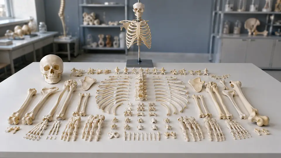
When the bones of a skeleton are joined into one standing figure, the body’s overall proportions are easy to see, but individual landmarks can disappear into the assembled form. A disarticulated skeleton model separates the bones so learners can examine them one at a time, compare neighboring structures, and reconstruct how the skeleton fits together. The term sounds technical, but the idea is straightforward: “disarticulated” means taken apart at the joints.
Instead of showing the femur locked into the hip or the radius and ulna positioned only as they appear in the forearm, this type of anatomy model presents bones as separate pieces. Depending on the design, the pieces may be loose, arranged on a support, or grouped by region. That separation changes the study experience from simply recognizing a skeleton’s outline to investigating the shape, surfaces, and relationships of its individual bones.
What Is a Disarticulated Skeleton Model?
A disarticulated skeleton model is a physical anatomical model in which the bones are separated from one another rather than permanently assembled into their normal anatomical positions. The word articulation refers to a joint or the meeting point between bones, so disarticulation means that those joint connections have been taken apart.
For example, on an assembled skeleton, the humerus sits between the scapula and the bones of the forearm. On a disarticulated version, the humerus can be handled and examined separately. Its rounded head, shaft, elbow surfaces, and bony projections are easier to distinguish when it is not surrounded by neighboring structures.
The same principle applies throughout the model. Vertebrae may be separated from one another, the ribs may be detached from the sternum, and the bones of the hand and foot may be presented as individual components. A complete set usually represents the major bones of the human skeleton, although the exact number of pieces and the inclusion of small bones vary by model.
How It Differs From an Articulated Skeleton
An articulated skeleton is assembled so that the bones are connected in, or close to, their natural anatomical relationships. Depending on the model, joints may be fixed, partially movable, or mounted on wires, springs, or other supports. This arrangement is useful for viewing posture, alignment, and the general organization of the skeleton.
A disarticulated model prioritizes separation instead. The learner can look closely at the irregular edge of a vertebra, rotate a scapula to inspect its surfaces, or place the bones of the forearm side by side. That freedom is particularly useful when the goal is to identify landmarks rather than simply observe the body as a whole.
| Feature | Disarticulated Skeleton | Articulated Skeleton |
|---|---|---|
| Bone arrangement | Bones are separated from one another | Bones are assembled in anatomical order |
| Main emphasis | Individual bone shape, landmarks, and identification | Overall alignment, posture, and joint relationships |
| Movement of pieces | Individual pieces can usually be repositioned | Movement depends on the model’s joint construction |
| Study activity | Sorting, identification, and reconstruction | Orientation, articulation, and regional demonstration |
| Storage | Requires organization for multiple separate pieces | Often remains assembled or mounted |
Neither format represents the “correct” skeleton by itself. A student studying the shoulder may prefer the loose scapula and humerus of a disarticulated set, while an instructor explaining how the shoulder girdle relates to the trunk may need an assembled display. The two designs answer different anatomical questions.
What Bones Are Included?
The contents depend on the manufacturer and the intended level of instruction. A full disarticulated skeleton commonly includes the skull, vertebral column, ribs, sternum, shoulder girdle, upper-limb bones, pelvic bones, and lower-limb bones. Some models also separate smaller elements, such as individual wrist and ankle bones, rather than grouping them into a single hand or foot piece.
Small bones make a noticeable difference during study. A student trying to learn the carpal bones, for instance, needs to distinguish several compact, similarly sized structures. A model that presents them separately creates an opportunity to arrange them in anatomical order and inspect their surfaces, although the pieces can also be easier to misplace.
Skull construction varies especially widely. Some disarticulated skeleton models include a skull that remains assembled, while others separate major skull bones or provide a removable mandible. These designs should not be treated as identical. A removable jaw demonstrates one kind of separation, whereas a fully disarticulated skull provides a much more detailed study of cranial bones and their interfaces.
Why Piece Count Can Be Misleading
Descriptions such as “22-part skull” or “multiple-piece skeleton” refer to the way a particular model is constructed, not to a universal anatomical standard. One model may count the left and right hip bones separately, while another may treat the pelvis as one assembled region. One may include paired small bones as separate pieces, while another may combine them for convenience.
For that reason, the written component list matters more than the number printed in a product title. When a lesson depends on a specific structure, such as the hyoid bone, individual vertebrae, or the ossicles of the ear, check whether that structure is actually provided as a separate component.
What Can You Study With a Disarticulated Model?
The strongest use of this model type is individual bone identification. A learner can examine the contours of the femur, locate its head and condyles, and then decide how the bone belongs in the body. The process is more active than reading a label beside a mounted skeleton because the student must use shape, side, and surface features together.
It also supports the study of anatomical landmarks. Ridges, fossae, foramina, processes, tubercles, and condyles are not decorative bumps. They provide attachment sites, passages, or joint surfaces, and their positions help identify a bone’s orientation. A separated pelvis, for example, can be turned in the hand so the acetabulum, iliac crest, and other landmarks are viewed from several angles rather than hidden by the rest of the skeleton.
Spatial relationships can be explored as well, although they require deliberate reconstruction. Placing the radius beside the ulna, fitting the vertebrae into a sequence, or arranging the carpal bones in the wrist turns the model into a three-dimensional sorting exercise. The table may become temporarily crowded with ivory-colored pieces, but that physical arrangement exposes relationships that a single flat image can compress.
Practical Study Activities
- Identify before labeling: Select a bone without looking at its name, then identify its region, side, major landmarks, and neighboring structures.
- Reconstruct a region: Arrange the bones of the forearm, hand, lower leg, or foot in anatomical order using an atlas or labeled reference for checking.
- Compare paired bones: Place the left and right versions together and note their mirrored features rather than treating them as interchangeable.
- Practice orientation: Hold a bone in anatomical position, then explain which surface is anterior, posterior, medial, or lateral.
- Use delayed recall: Study a labeled diagram, set it aside, and test identification using only the physical pieces.
These exercises do not make the model a replacement for an atlas, lecture, or digital resource. A reference image can supply labels and terminology, while the model supplies shape and depth. Used together, those tools allow a learner to move between names on a page and structures that occupy real three-dimensional space.
Disarticulated Does Not Always Mean Completely Loose
The word “disarticulated” describes the relationship between the bones, not necessarily the storage method. Some models contain entirely separate pieces in a case or tray. Others use a stand, connecting rods, magnets, wires, or regional assemblies to keep components organized while still presenting the bones apart from their joints.
A model may therefore be disarticulated in one area and assembled in another. For example, the limbs might be separated while the vertebral column remains threaded on a support. A skull may stay intact even though the rest of the skeleton is supplied as individual bones. This mixed construction is not automatically a contradiction, but the specifications should be read carefully.
There is also a difference between a disarticulated model and a model with removable parts. A skeleton with a removable arm may still be fundamentally articulated. The arm has been detached from the rest of the display, but its own bones may remain connected in anatomical position. Disarticulation applies to the joints between the bones themselves.
Advantages and Limitations for Learning
Separation provides excellent access to bone morphology. A learner can rotate a piece, view its underside, compare it with a similar bone, and inspect areas that would be partly hidden in an assembled skeleton. This is particularly valuable for the irregular bones of the vertebral column, pelvis, skull, wrist, and ankle.
The format also encourages active handling and reconstruction. Instead of passively following a label, the student can ask, “Which way does this face?” or “What fits beside this surface?” Those questions bring orientation and relationships into the lesson, where simply memorizing a list of names may leave gaps.
There are practical limitations. Loose pieces can be misplaced, and small bones may be difficult to manage in a busy classroom. A disarticulated set also does not show posture or full-body alignment as immediately as a mounted skeleton. A learner who studies only separate bones may know the landmarks yet struggle to visualize how the pelvis, spine, and lower limbs relate in the standing body.
For that reason, many anatomy courses use both arrangements. An articulated skeleton supplies the body-wide framework, while a disarticulated set supports close inspection and self-testing. Even a labeled atlas or 3D digital model can fill gaps by showing structures from viewpoints that are difficult to reproduce with loose physical pieces.
Care, Organization, and Storage
Care requirements depend on the model’s plastic, paint, labels, fasteners, magnets, and support hardware. Follow the manufacturer’s instructions when they are available, especially before applying any cleaner. A soft, dry or slightly damp cloth is often safer for routine dust removal than abrasive materials, but the model’s specific construction should determine the final method.
Organization matters as much as cleaning. Small trays, labeled compartments, or a parts inventory can keep carpals, tarsals, ribs, and vertebrae from becoming a confusing pile. After a practical session, returning each piece to its designated location reduces the chance that a missing bone will be mistaken for an incomplete model.
Handle thin projections and small bones carefully, and avoid forcing pieces into supports that are not designed for them. Printed labels can wear from repeated rubbing, while adhesives and painted surfaces may react poorly to strong solvents. A cool, dry storage area away from prolonged direct sunlight is generally sensible, but any special material warnings should come from the manufacturer.
Frequently Asked Questions
Is a disarticulated skeleton model made from real human bones?
Educational disarticulated skeleton models are typically manufactured replicas rather than real human skeletal remains. They are commonly produced from molded materials and may include painted or printed details. The exact material and finish vary by model, so product documentation is the appropriate source for construction details.
Can a disarticulated skeleton be put back together?
Sometimes. If the model includes separate pieces without a permanent mounting system, learners can arrange the bones into anatomical order. However, a complete reconstruction may require a reference image, and some models are designed primarily for examination rather than permanent assembly.
Is a disarticulated model better than an articulated skeleton?
Neither is universally better. A disarticulated model is especially useful for individual bone identification, landmarks, and reconstruction exercises. An articulated skeleton is more convenient for studying posture, alignment, and the relationships of bones across the whole body.
What is the difference between a disarticulated skeleton and a skeleton with removable limbs?
A disarticulated skeleton separates bones at their joints. A skeleton with removable limbs may only allow an entire arm or leg to detach from the main frame, while the bones within that limb remain articulated. The distinction is the level at which separation occurs.
The Practical Meaning of the Term
In plain language, a disarticulated skeleton model is a skeleton presented as separate bones rather than one connected figure. Its purpose is not simply to show that the body contains bones. It allows learners to study each bone’s form, orientation, landmarks, and relationship to its neighbors through sorting, comparison, and reconstruction.
When evaluating a description, look beyond the word “disarticulated.” Check which bones are actually separate, whether the skull is divided, how small bones are stored, and whether any regions remain assembled on a support. Those details determine what the model can demonstrate and how it will function during study.
