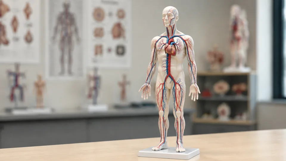
Vascular anatomy is as much about pathways and relationships as it is about naming individual vessels. The aorta, basilar artery, pulmonary trunk, and coronary branches become easier to distinguish when their courses can be followed through three-dimensional space rather than viewed only on a page. Vascular anatomy models make those relationships visible, ranging from focused cerebral vessel networks to broader circulatory-system displays designed for study, teaching, and demonstration..
The most useful model depends on the anatomical system or teaching task being studied. A neurology student may benefit from a detailed cerebral vessel model, whereas a broader circulatory model can provide better context for tracing vessels through the chest and major organs. Scale, color coding, removable components, reference materials, base stability, and viewing distance all influence how effectively a model works for individual study or classroom demonstration.
For broad cardiopulmonary instruction, the MediTrainer Human Cardiopulmonary Vascular Anatomy Model is the strongest overall option here. Its life-size format, heart and lung vessels, removable display arrangement, and labeled reference chart create a practical balance between detail and usability. Still, cerebral circulation, coronary anatomy, systemic pathways, and disease education each call for a different kind of model.
Quick Answer
Best overall: The MediTrainer Human Cardiopulmonary Vascular Anatomy Model combines a life-size heart-lung presentation with color-coded vessels and a supporting reference chart. Best broad circulatory display: The Half Life Size Human Circulatory System Anatomy Relief Model places major vessels beside the heart, lungs, liver, kidneys, and other organs. Best for cerebral arteries and veins: The MediTrainer Human Brain Arteriovenous Vascular Model displays both networks in contrasting colors. For straightforward public-health demonstrations, the compact Simedutech Atherosclerosis Cardiovascular Model makes arterial narrowing easy to see across four stages.
For a fast side-by-side view, the table below highlights the anatomical focus and most useful teaching features of all nine selections.
Best Vascular Anatomy Models Comparison Table
| Product | Best For | Scale or Format | Key Feature |
|---|---|---|---|
| MediTrainer Cardiopulmonary Vascular Model | Heart-lung circulation | Life-size | Color-coded pulmonary vessels and internal heart structures |
| MediTrainer Brain Arteriovenous Model | Cerebral arteries and veins | Detachable display model | Complete red and blue cerebral vessel networks |
| MediTrainer Brain Artery Model | Basilar artery study | Life-size | Named basilar branches and brainstem structures |
| Half Life Size Circulatory System Relief Model | Classroom-wide instruction | Half-size 3D relief | Systemic vessels shown beside major organs |
| Merinden Respiratory System Model | Removable cardiopulmonary study | Life-size, seven pieces | Detachable lungs, heart sections, and throat components |
| Simedutech Atherosclerosis Model | Vascular disease education | Compact tabletop model | Four stages of arterial plaque progression |
| EVOTECH SCIENTIFIC Urogenital Model | Renal and pelvic vessels | Life-size, three parts | Kidneys, ureters, bladder, arteries, and veins |
| qzzXoq Head and Neck Model | Head and neck relationships | Head, neck, and chest display | Vascular and neural structures beside layered muscles |
| NEW HORIZON Vascular Heart Model | Coronary circulation | Natural size | Prominent coronary vessels and two stents |
The full reviews explain where each model shines, what it leaves out, and which learning environment suits it best.
1. MediTrainer Human Cardiopulmonary Vascular Anatomy Model: Best Overall

This life-size MediTrainer model makes the heart-lung circuit feel concrete rather than abstract. Red arterial branches spread through the lungs while blue and purple vessels establish a clear visual distinction between pathways. Internal heart structures add context, letting an instructor move from the pulmonary trunk and pulmonary veins into the atria and ventricles without swapping between several teaching aids.
The metal support and smooth base keep the branching assembly elevated for display, yet the model can be removed from its bracket for a closer view. Picture a nursing instructor passing it along the front row while students follow a vessel toward the heart, then returning it to the stand without a wobbly balancing act. A labeled chart identifies structures such as the aorta, coronary artery, pulmonary trunk, and heart chambers, which is especially useful during self-study.
Pros
- Life-size presentation provides strong spatial context.
- Color-coded vessels distinguish major circulatory pathways clearly.
- Shows pulmonary vasculature alongside internal heart anatomy.
- Detaches from the support for close observation.
- Includes a labeled anatomical reference chart.
Cons
- Its specialized heart-lung focus does not cover systemic circulation throughout the body.
- Fine pulmonary branches require careful handling during frequent classroom use.
This is the most balanced selection for cardiopulmonary courses, nursing instruction, respiratory education, and clinical explanations centered on blood flow between the heart and lungs. Buyers needing cerebral vessels or a whole-body overview should choose a more specialized option below. For a desk or demonstration table, however, its combination of recognizable scale, branching detail, and stable presentation is hard to beat.
To buy this product, click here.
2. MediTrainer Human Brain Arteriovenous Vascular Model: Best for Cerebral Arteries and Veins

Cerebral vasculature can look like a tangled bowl of noodles in a textbook illustration. This MediTrainer model separates the networks with red arteries and blue veins, helping learners follow their distribution through three-dimensional space. That dual-network view sets it apart from artery-focused models and makes it useful for broader cerebral circulation lessons.
The vascular assembly sits on a smooth wooden base, creating a polished display for a neurology classroom, laboratory, or consultation desk. It can also be detached for closer inspection, a welcome feature when students need to turn the model and inspect branches hidden from a frontal view. The included manual labels cerebral arteries and veins and explains circulation, providing a road map through an otherwise visually dense structure.
Pros
- Displays cerebral arterial and venous networks together.
- Red and blue coloring supports rapid vessel identification.
- Detachable structure can be viewed from multiple angles.
- Wooden base provides a stable, presentable display.
- Includes a detailed vessel reference manual.
Cons
- The intricate exposed branches call for gentler handling than a solid organ model.
- It is less focused than an artery-only model for isolated basilar artery lessons.
Neurology students, nursing programs, and educators seeking a full cerebral vessel network should place this near the top of the list. Its chief advantage is the simultaneous arterial and venous view. If the lesson concentrates on named basilar branches and their relationship to the brainstem, the next model offers a narrower and potentially clearer presentation.
To buy this product, click here.
3. MediTrainer Brain Artery Model: Best for Basilar Artery Study
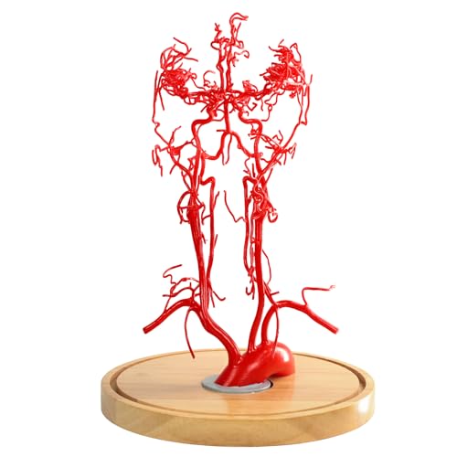
For learners concentrating on posterior cerebral circulation, this life-size model takes a more targeted route. It presents the basilar artery and branches that include the superior cerebellar, anterior inferior cerebellar, and posterior inferior cerebellar arteries. Those names can blur together during a late-night review session, but seeing each branch occupy physical space gives the terminology somewhere to stick.
Brainstem landmarks, including the pons and medulla oblongata, provide essential orientation. Rather than viewing arteries as floating red lines, students can study how branches relate to nearby neural anatomy. The PVC construction is described as odorless and easy to clean, making the model suitable for repeated handling in teaching spaces where markers, notebooks, and coffee cups tend to crowd every available surface.
Pros
- Focused depiction of the basilar artery and major branches.
- Includes pons and medulla oblongata for spatial context.
- Life-size format supports realistic anatomical orientation.
- PVC surface is intended for repeated use and cleaning.
- Well suited to neurology and neuroscience instruction.
Cons
- Does not provide the same complete artery-and-vein comparison as the arteriovenous pick.
- Its specialized scope has less value for general systemic circulation courses.
This model makes sense for educators who need to point to a specific cerebellar branch without hunting through a crowded full-brain network. It also complements brainstem coursework nicely. General anatomy students seeking one broad vascular model may get greater mileage from the cardiopulmonary or circulatory relief choices, while focused neuroscience learners will appreciate this model’s disciplined scope.
To buy this product, click here.
4. Half Life Size Human Circulatory System Anatomy Relief Model: Best for Classrooms

A focused organ model excels up close, but a room full of students often needs a broader visual anchor. This half-natural-size relief presents arteries and veins alongside the heart, lungs, liver, spleen, kidneys, skeletal sections, and other anatomical landmarks. The raised format creates more depth than a flat poster while remaining easier to display than a freestanding full-body vascular network.
Red and blue coloring allows an instructor to trace broad pathways from several feet away. Imagine a biology teacher standing beside the display, moving from the heart toward renal vessels and back to the lungs while students sketch the route. The PVC relief is lightweight and easy to clean, and the listing describes it as suitable for wall or tabletop presentation, giving crowded classrooms some welcome flexibility.
Pros
- Provides the broadest systemic overview among these selections.
- Places major vessels in context with multiple organs.
- Half-size relief is visible during group demonstrations.
- Red and blue coloring distinguishes arteries and veins.
- Supports wall or tabletop educational display.
Cons
- Relief construction offers less all-angle exploration than a freestanding vessel model.
- Its broad scope provides less fine detail for individual cerebral or coronary branches.
Choose this model for introductory anatomy, secondary-school biology, patient education, or classrooms where a clear systemic map matters more than tiny vessel landmarks. It makes relationships between organ systems easy to discuss at a glance. Advanced learners studying one vascular territory should pair that broad perspective with a focused model, but instructors seeking one prominent room display will find this format especially practical.
To buy this product, click here.
5. Merinden Life-Size Human Respiratory System Anatomy Model: Best Removable Design

The Merinden model approaches cardiopulmonary anatomy as a layered puzzle. Its seven pieces include two throat sections, two lungs, two heart sections, and a base with the trachea. Arteries, veins, the aorta, and pulmonary arteries appear within that larger respiratory presentation, allowing students to explore how airways, lungs, heart, and vessels occupy the same compact region.
Removable components make this particularly useful for practical teaching. An instructor can lift away a lung, open the heart sections, and shift from surface recognition to internal orientation during one lesson. Numbered structures support identification, while the included chart supplies a reference when the model is used for independent review. Once reassembled, the sturdy base keeps the seven-part model organized for display.
Pros
- Seven removable pieces encourage hands-on anatomical study.
- Life-size format gives the structures realistic context.
- Combines respiratory organs with major cardiopulmonary vessels.
- Numbered structures simplify guided identification exercises.
- Includes a sturdy base and explanatory chart.
Cons
- Respiratory anatomy receives more emphasis than fine vascular branching.
- Multiple removable pieces require more setup and organization than a fixed display.
This is a strong fit for anatomy laboratories, respiratory programs, and instructors who want students to disassemble structures rather than stare at a fixed piece. Buyers interested purely in vessel networks may find the top MediTrainer option more direct. Those who teach how circulation and respiration intersect, however, gain a versatile model that can support several lessons instead of one narrow demonstration.
To buy this product, click here.
6. Simedutech Human Atherosclerosis Cardiovascular Model: Best for Disease Education

Sometimes a small, focused model lands the lesson harder than a sprawling network. Simedutech depicts four steps in atherosclerosis progression: a normal artery, fatty streak, fibrous plaque, and occlusive plaque. Placed side by side, the stages create an immediate visual comparison of how an open lumen changes as plaque develops, a concept that can feel oddly flat on a printed page.
The model measures 7.4 by 5.8 by 3.3 inches, so it fits comfortably on a consultation desk, health-fair table, or lecture podium. Textured vascular surfaces and lifelike coloring help differentiate the stages, while the base keeps everything upright during discussion. An information card is included, giving educators a compact reference for public-health presentations and introductory cardiovascular lessons.
Pros
- Shows four clearly differentiated stages of atherosclerosis.
- Compact footprint suits desks and portable demonstrations.
- Progressive layout communicates arterial narrowing quickly.
- Stable base supports frequent educational display.
- Includes an information card for guided explanation.
Cons
- Focuses on disease progression rather than the wider vascular network.
- Small format is less visible than a large relief model in spacious classrooms.
Health educators, clinics, and instructors introducing vascular disease will get the clearest value from this model. It is simple enough for a brief conversation yet structured enough for students to compare arterial stages. Anyone studying named vessels or regional circulation should look elsewhere, since this is a concept model rather than a vascular map. For one sharp lesson about lumen narrowing, its visual economy is precisely the appeal.
To buy this product, click here.
7. EVOTECH SCIENTIFIC Male Urogenital System Anatomical Model: Best for Renal and Pelvic Vessels

Regional anatomy makes more sense when vessels remain attached to the organs they serve. This life-size EVOTECH SCIENTIFIC model depicts the kidneys, adrenal glands, ureters, bladder, arteries, and veins within the male urinary and reproductive system. That arrangement suits lessons where renal and pelvic circulation need to be discussed alongside urinary structures rather than isolated as floating branches.
Three removable parts give learners access to different viewing angles and make the model more interactive than a fixed regional display. The PVC body is lightweight and easy to clean, while a sturdy base keeps the arrangement stable between demonstrations. Its instruction manual contains labeled diagrams, useful when a student is working alone and needs to match a molded structure with the correct name.
Pros
- Shows arteries and veins within realistic urogenital context.
- Life-size presentation supports regional orientation.
- Three-part construction permits closer examination.
- Stable base suits classroom and laboratory display.
- Includes a manual with labeled anatomical diagrams.
Cons
- Vascular anatomy is secondary to the broader urogenital-system presentation.
- Its male-specific regional focus limits usefulness for general circulation instruction.
This model is a sensible choice for renal, urinary, and male reproductive anatomy modules where vascular relationships form one part of a larger lesson. It is far less appropriate as a first general-purpose vascular model, since its vessels remain confined to a specific anatomical region. For targeted coursework, that narrow context becomes helpful rather than restrictive, especially during organ-identification exercises.
To buy this product, click here.
8. qzzXoq Head and Neck Chest Muscle Anatomical Model: Best for Head and Neck Relationships

The head and neck pack vessels, nerves, and layered muscles into an extraordinarily tight space. This qzzXoq model emphasizes those relationships across the face, neck, and upper chest, displaying superficial and deeper musculature with vascular and neural structures in context. For learners who keep losing track of what passes beside, beneath, or across a muscle, that three-dimensional neighborhood is the core attraction.
Numbered landmarks support structured study and allow an instructor to call out features during a lab or demonstration. The included manual helps decode those identifiers rather than leaving students to guess at a dense field of colored structures. Its odorless, corrosion-resistant PVC construction is intended for repeated educational use, and the model can double as a prominent laboratory reference between lessons.
Pros
- Integrates facial and neck vessels with nerves and muscles.
- Shows superficial and deeper muscular relationships.
- Numbered landmarks support guided identification.
- Includes a manual for anatomical reference.
- Relevant to head, neck, and maxillofacial education.
Cons
- Dense muscular and neural detail can feel busy for beginners studying vessels alone.
- Provides less dedicated vascular detail than a pure cerebral vessel model.
Advanced head and neck students, maxillofacial learners, and educators teaching regional relationships are the natural audience. This is a context-rich model rather than a clean vascular map, so its visual density is purposeful. Introductory students who simply need to distinguish arteries from veins may prefer a clearer color-coded circulatory display, while regional learners can benefit from seeing how crowded this anatomy truly is.
To buy this product, click here.
9. NEW HORIZON Human Vascular Heart Model: Best for Coronary Circulation

Coronary vessels deserve more attention than they receive on many general heart models. This natural-size NEW HORIZON model emphasizes cardiac veins and coronary arteries wrapping around the myocardium, creating a direct view of the vessels responsible for supplying and draining heart muscle. The enhanced coronary detail is especially handy when a standard red-and-blue heart begins to feel too simplified.
Two inserted stents add another concrete teaching feature. During a cardiology lesson or patient-oriented explanation, an educator can point to their placement while discussing coronary anatomy and arterial blockage in general educational terms. The stable base keeps the model ready on a desk, and its focused exterior arrangement avoids the visual clutter of a complete cardiopulmonary network.
Pros
- Emphasizes coronary arteries and cardiac veins.
- Natural-size heart provides recognizable anatomical context.
- Includes two visible stents for educational demonstration.
- Enhanced coronary pathways are easier to trace than on simpler heart models.
- Mounted base supports convenient tabletop display.
Cons
- Exterior coronary emphasis provides less internal heart detail than a sectional model.
- Its cardiac focus does not illustrate pulmonary or systemic circulation comprehensively.
Cardiology students, educators, and clinics seeking a focused coronary display should consider this over a generic heart model. It is particularly useful when the lesson centers on myocardial blood supply, cardiac veins, or the location of coronary stents. Buyers needing chambers, valves, lungs, and pulmonary vessels in one presentation will gain more from the overall winner, but this model keeps coronary anatomy front and center.
To buy this product, click here.
How to Choose a Vascular Anatomy Model
Start With the Anatomical Region
Begin by naming the lesson rather than choosing the most dramatic model on the shelf. Cardiopulmonary circulation calls for visible heart-lung connections, cerebral instruction needs intracranial branches, and a renal lesson benefits from vessels shown beside the kidneys and ureters. A medical student reviewing the basilar artery at midnight has little use for a broad torso display, while a high-school teacher introducing systemic circulation may find that same brain model far too specialized.
Match Detail to the Learner
More detail can help advanced students, but it can bury a beginner beneath a thicket of branches. Clean red and blue pathways work well for introductory identification and room-wide demonstrations. Named branches, neural landmarks, or dense regional relationships are better suited to learners who already understand basic circulation. Think about the viewing distance too. Tiny coronary features may look crisp across a desk and disappear from the back row.
Check Scale and Display Format
Life-size models provide intuitive spatial context, especially for the heart, lungs, brain, and regional organs. Half-size reliefs trade full scale for a wider anatomical field and easier wall display. Compact disease models sacrifice geographic detail but travel neatly to health fairs and consultation rooms. Before ordering, picture the actual space: a narrow laboratory shelf, a mobile teaching cart, or a permanent classroom wall each favors a different format.
Decide Whether Removable Parts Matter
Detachable lungs, split heart sections, and lift-off vascular assemblies invite close inspection and hands-on lessons. They also create pieces that must be tracked, assembled correctly, and handled with care. Fixed relief models are quicker to present and harder to misplace. If several student groups will rotate through a station, removable components can be engaging, provided the course benefits from seeing obscured surfaces or internal structures.
Look for Useful Color Coding and References
Red and blue coloring helps learners distinguish arterial and venous systems quickly, though color alone does not explain oxygenation in every vessel. Pulmonary arteries and veins are a classic reason instructors must reinforce names, direction of flow, and function rather than relying solely on color. Manuals, numbered landmarks, and labeled charts reduce friction during independent study, particularly when dozens of similar branches crowd the view.
Consider the Base and Handling Demands
Branching vascular models can be delicate because their educational value comes from exposed, slender structures. A stable base protects the model from casual knocks, and a detachable stand makes close viewing easier. Smooth PVC surfaces are practical for repeated classroom use, but intricate branches still deserve careful storage. A sturdy relief may be the calmer choice in a busy room where backpacks bump tables and models move between classes.
Best Vascular Anatomy Models FAQ
Which Vascular Anatomy Model Is Best for Medical Students?
The best choice follows the course module. The MediTrainer Cardiopulmonary Vascular Model offers a useful general foundation for heart-lung circulation, while neurology students may prefer one of the cerebral models. Students studying several systems may benefit from a broad circulatory relief first, followed by a focused regional model as coursework becomes more detailed.
Are Life-Size Models Better Than Reduced-Scale Models?
Life-size models make distances and organ relationships easier to interpret, but reduced-scale displays can cover more of the body without consuming an entire room. A half-size relief works well for group teaching because it presents numerous organs and vessels together. Life-size regional models are usually more useful for close desk study and detailed demonstrations.
Should a Vascular Model Show Both Arteries and Veins?
Choose both networks for lessons about circulation, vessel comparison, or venous drainage. An artery-only model can be clearer when studying a specific arterial territory such as the basilar branches. The teaching objective should decide whether additional structures provide context or create visual clutter.
Are Removable Anatomy Models Better for Learning?
Removable parts help learners inspect hidden surfaces and understand how structures fit together. They are particularly useful in laboratory stations and small-group demonstrations. Fixed models remain valuable for quick reference, wall display, and busy classrooms where loose components could be misplaced.
What Makes a Model Suitable for Patient Education?
Clear color contrast, a stable base, moderate detail, and an obvious visual lesson matter most. The Simedutech model communicates plaque progression quickly, while a cardiopulmonary model can support general explanations of heart-lung circulation. These products are educational aids and do not replace individualized explanations from qualified healthcare professionals.
Can an Anatomy Model Replace an Atlas or Textbook?
No. A model supplies spatial context, while an atlas or textbook provides terminology, variation, function, and deeper explanation. The strongest study setup uses both. Trace a pathway on the model, identify it through the included chart or manual, then confirm the details in course materials.
How Should Intricate Vascular Models Be Stored?
Keep branching models on their supplied bases in a low-traffic location, away from shelf edges and heavy objects. Lift them by the main support or base rather than a vessel branch. Removable pieces should stay together in a labeled container if the model will be transported between rooms.
Final Verdict
The MediTrainer Human Cardiopulmonary Vascular Anatomy Model is the strongest overall pick among these nine models. Its life-size heart-lung arrangement, contrasting vessel colors, internal cardiac detail, removable support system, and labeled chart cover the features most buyers need for cardiopulmonary education. It is focused enough to remain readable yet broad enough to support anatomy courses, nursing instruction, and visual explanations around a desk or classroom table.
Different lessons deserve different tools. Choose the Half Life Size Human Circulatory System Anatomy Relief Model for a broad classroom overview, the MediTrainer Human Brain Arteriovenous Vascular Model for cerebral arteries and veins, or the compact Simedutech Atherosclerosis Cardiovascular Model for a clear progression of arterial plaque. Before selecting one, write down the target region, learner level, viewing distance, and need for removable parts. That short checklist will steer you toward a model that teaches the intended anatomy clearly instead of becoming an impressive but mismatched shelf ornament.
