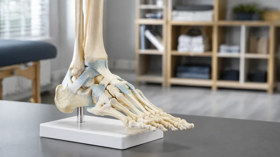
Twenty-six bones, dozens of joints, and a dense web of ligaments fit into a remarkably small space beneath the ankle. That compact architecture is exactly why a screen diagram can feel clear at breakfast and hopelessly tangled by exam time. A physical ankle anatomy model lets students rotate the calcaneus, trace the metatarsals, and see how the tibia and fibula meet the talus in three dimensions. In a clinic or classroom, the same model can turn a fuzzy explanation into a quick, concrete demonstration.
Models in this category vary sharply. Some focus on articulated bones, while others add ligaments, muscle attachments, nerves, vessels, or pathology. Buyers often pick a life-size skeleton without checking which side it represents, whether it leaves the stand, or how much movement the wiring permits. The most useful choice depends on the job. Students may favor clear landmarks and portability, instructors may need sturdy articulation, and podiatry educators may require layered plantar anatomy.
The Evotech Scientific Human Foot and Ankle Model is the strongest all-around pick for accessible bone study, thanks to its life-size proportions, flexible construction, removable stand, and detailed right-foot skeleton. More advanced learners should look closely at the Axis Scientific 9-Part Model, whose removable muscles, ligaments, nerves, and arteries reveal far more than a basic bone replica.
Quick Answer
Best overall: The Evotech Scientific Human Foot and Ankle Model offers a practical balance of anatomical detail, articulation, and desk-friendly display. Best detailed option: The Axis Scientific 9-Part Foot Anatomy Model suits layered study of muscles, ligaments, nerves, vessels, and plantar structures. Best for patient education: The GPI Anatomicals model makes plantar fasciitis and major ankle structures easy to point out. Best for muscle attachments: The painted Merinden model highlights origin and insertion areas in red.
For a fast side-by-side view, the table below compares all nine selections before the full reviews.
Best Ankle Anatomy Models Comparison Table
| Product | Best For | Anatomical Detail | Movement or Construction |
|---|---|---|---|
| Evotech Scientific Foot and Ankle Model | Overall bone study | Right foot, tibia, fibula, and foot bones | Flexible wire and elastic ankle connection |
| Axis Scientific 9-Part Foot Model | Layered anatomy | Muscles, ligaments, nerves, arteries, and plantar anatomy | Nine removable components |
| GPI Anatomicals Foot and Ankle Model | Patient education | Major bones, ankle ligaments, Achilles tendon, and plantar fascia | Fixed display model with base |
| Merinden Painted Foot and Ankle Model | Muscle attachments | Right-foot skeleton with red attachment markings | Fully articulated |
| QWORK Foot Skeleton Model | Students on a budget | Life-size right foot with lower-leg bone segments | Articulated and detachable from base |
| Human Right Foot Bone Anatomy Model | Ligament visualization | Twenty-six foot bones with ligament representation | Flexible ankle, midfoot, and toe joints |
| NEW HORIZON Foot Ankle Model | Art and anatomy reference | Right foot, tibia, fibula, and textured bone landmarks | Flexible wire, elastic connection, removable base |
| Human Foot Ankle Model 1:1 | Left-foot study | Left foot with tibia and fibula segments | Stainless-steel wire articulation |
| qzzXoq Six-Piece Joint Model Set | Whole-body joint teaching | Foot plus hand, hip, knee, shoulder, and elbow | Flexible ligament-equipped joint models |
The closer reviews explain where each model shines and which anatomical compromises matter during study or demonstration.
1. Evotech Scientific Human Foot and Ankle Model — Best Overall

This Evotech Scientific model covers the fundamentals without turning a study desk into a crowded laboratory bench. The listing describes a life-size right foot cast from a human specimen, with the tibia, fibula, tarsals, metatarsals, and phalanges represented. Textured PVC surfaces make ridges and joint contours easier to follow than they appear in a glossy illustration. Picture a student rotating the heel before a practical exam, tracing from the talus through the navicular and into the forefoot. That tactile sequence is where this model earns its top ranking.
Flexible stainless-steel wire holds the foot and toe bones together, while an elastic connection at the ankle permits demonstrative movement. It will not reproduce the controlled biomechanics of a living foot, yet it offers enough motion to show how the skeletal sections relate. The model also lifts away from its compact 4.3-inch-square base, which is useful when several students need a close view. Set it upright for display, then pull it free to inspect the sole, heel, or ankle mortise from a better angle.
Pros
- Life-size right-foot skeleton includes tibia and fibula segments
- Specimen-based casting preserves useful bone contours
- Flexible wire supports ankle, foot, and toe movement
- Detaches from the stand for closer inspection
- Easy-clean PVC suits repeated classroom use
Cons
- Does not include color-coded muscles, nerves, or vessels
- Flexible articulation demonstrates relationships rather than precise physiological range
This is a sensible choice for anatomy students, artists, physical therapy trainees, and educators who primarily need skeletal landmarks. Buyers teaching soft-tissue anatomy will outgrow it faster than the Axis model, but its uncluttered bones are actually helpful during early identification work. For a backpack-to-desk model that can be handled, turned, and displayed with little fuss, it hits a particularly useful sweet spot.
To buy this product, click here.
2. Axis Scientific 9-Part Foot Anatomy Model — Best Detailed Option
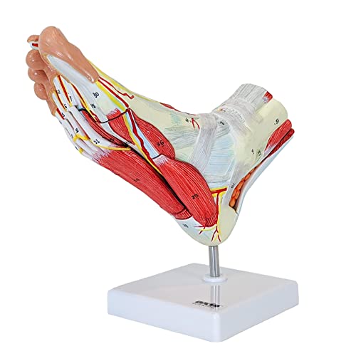
Peeling anatomy back layer by layer has a satisfying clarity that a bare skeleton cannot match. This life-size Axis Scientific model separates into nine components, exposing muscles, ligaments, nerves, and arteries as pieces come away. A podiatry student can begin with the assembled foot, remove superficial structures, and then connect deeper anatomy to the visible surface. That progression makes dense spatial relationships feel far less abstract.
The plantar surface is the headline feature. Many models lavish attention on the top of the foot while leaving the sole sparse, yet this one represents nerves and arteries running beneath it. That makes it useful for discussing nerve pathways, musculotendinous relationships, ligament injuries, and plantar structures in an educational setting. A full-color study guide uses photographs of the model with numbered structures identified, and the product includes a three-year warranty according to the listing. It carries more visual information than beginners may need, but serious learners will appreciate the depth.
Pros
- Nine removable components support progressive layer-by-layer study
- Represents muscles, ligaments, nerves, and arteries
- Includes detailed anatomy beneath the sole
- Life-size format works well for close teaching and patient explanation
- Includes a photographic study guide and listed three-year warranty
Cons
- More complex than a basic skeleton for first-pass bone identification
- Multiple removable pieces require careful storage and organization
Graduate-level health science students, podiatry educators, sports medicine programs, and clinics are the clearest audience. Someone memorizing the tarsals for an introductory course can save space and money with the Evotech or QWORK models. For teaching how vessels, nerves, muscles, and ligaments occupy the same tight anatomical landscape, however, the Axis model is the standout of these nine picks.
To buy this product, click here.
3. GPI Anatomicals Foot and Ankle Model With Plantar Fasciitis — Best for Patient Education

A patient hearing “plantar aponeurosis” may picture almost anything. Pointing to the band on a three-dimensional foot is quicker and much less foggy. The GPI Anatomicals replica presents plantar fasciitis alongside major skeletal and connective structures, making it particularly useful on a clinic desk. Its anatomy includes the tibia, fibula, calcaneus, Achilles tendon, deltoid ligament, lateral collateral ligament, plantar aponeurosis, and principal foot bones.
The model measures 9 by 2.75 by 4 inches and sits on a 6.5-by-5-inch display base, so it remains visible without swallowing a reception counter or teaching podium. An information card accompanies the display and gives an instructor another reference during conversation. Unlike a loose articulated skeleton that bends and rattles in the hand, this format emphasizes immediate visual explanation. A clinician can point from heel to plantar fascia while the model stays planted on the desk, which is often exactly what a short educational conversation needs.
Pros
- Illustrates plantar fasciitis in a clear physical format
- Displays major ankle ligaments and the Achilles tendon
- Includes tibia, fibula, calcaneus, and principal foot bones
- Stable display base suits clinics and teaching spaces
- Comes with a compact informational card
Cons
- Less suitable for learners who want to manipulate individual joints
- Focus on selected structures provides less layered detail than a removable-part model
This GPI model fits podiatry offices, physical therapy teaching spaces, and classrooms discussing common heel and plantar anatomy. Students seeking extensive articulation should move toward the skeletal picks, while advanced learners studying nerves and vessels should choose Axis Scientific. Its real strength is communication: the relevant anatomy remains exposed, steady, and ready for a quick point-and-explain moment.
To buy this product, click here.
4. Merinden Painted Life-Size Human Foot and Ankle Model — Best for Muscle Attachments

Red attachment markings change the way a plain skeleton reads. Rather than staring at an ivory surface and trying to recall where a muscle anchors, learners get an immediate visual cue on this Merinden right-foot model. The life-size skeleton includes tibia and fibula segments, helping students connect the ankle to the lower leg instead of treating the foot as an isolated object. For biomechanics coursework, those proximal connections matter.
Full articulation adds another layer of usefulness. An instructor describing gait can move the model while pointing out how the ankle and foot structures align, and a student can inspect the underside without losing the broader orientation. The stable base keeps the piece upright between demonstrations, which saves it from rolling around a crowded lab table beside notebooks and coffee cups. This is still a skeleton rather than a removable muscle model, so the red markings indicate attachment areas rather than showing complete muscles in three dimensions.
Pros
- Red markings identify muscle origin and insertion areas
- Life-size proportions support direct spatial comparison
- Includes tibia and fibula segments for ankle context
- Articulated construction supports movement demonstrations
- Stable base keeps the model accessible for display
Cons
- Attachment markings do not replace three-dimensional muscle structures
- Color cues may feel visually busy during bone-only identification practice
Kinesiology students, massage therapy learners, artists, and instructors covering muscle attachments should place this high on the list. A clean unpainted skeleton is calmer for early osteology drills, while Axis Scientific offers greater soft-tissue depth. Merinden occupies the useful middle ground, bringing muscular context to an articulated skeleton without requiring users to manage a collection of removable pieces.
To buy this product, click here.
5. QWORK Human Foot Skeleton Model — Best Value for Students

For a student who needs to turn flash-card knowledge into a mental map, QWORK offers a straightforward life-size right-foot skeleton. It represents the fibula, tibia, tarsals, metatarsals, and phalanges in corrosion-resistant PVC. The uncluttered presentation works well for repeated identification sessions. Place a finger on the talus, follow the arch, then rotate the toes and repeat until the sequence finally sticks.
The articulated design permits flexible foot movement, and the model can be detached from its base for closer observation. That removable setup matters more than it sounds. A mounted model looks tidy during a lecture, but a learner often needs to flip it over, peer between bones, and view the ankle from the side. The listing also refers to ligament and tendon representation, adding context beyond the bone shapes. Its modest, practical construction targets daily study rather than an elaborate layered dissection experience.
Pros
- Life-size right-foot format supports basic anatomy study
- Includes tibia and fibula segments
- Flexible articulation demonstrates skeletal relationships
- Detaches from the base for close viewing
- Easy-clean PVC is practical for classrooms
Cons
- Lacks the removable muscles and neurovascular detail of advanced models
- Broad articulation is better for demonstration than precise range-of-motion analysis
QWORK makes sense for nursing students, introductory anatomy courses, artists, and home study areas where affordability and handling matter most. Buyers seeking pathology, color coding, or layered plantar anatomy should spend their budget elsewhere. For learning the names, positions, and basic relationships of foot and ankle bones, its simple approach keeps attention where it belongs.
To buy this product, click here.
6. Human Right Foot Bone Anatomy Model With Ligaments — Best for Ligament Visualization

Ligaments are the feature that separates this model from the many bare-bone options clustered around the same category. The life-size right foot represents all 26 foot bones across the hindfoot, midfoot, and forefoot, including the talus, calcaneus, tarsals, metatarsals, and phalanges. Its ligament representation helps explain why neighboring bones hold together, a relationship that can disappear when learners study isolated labels on a page.
The listing describes integrated molding with textured ridges, attachment points, and joint surfaces. Flexible articulation extends across the ankle, midfoot, and toes, which gives an instructor several regions to manipulate during a demonstration. Imagine explaining inversion and eversion while students crowd around a desk. A movable physical model makes the direction of travel easier to see, though its flexibility should be treated as an educational illustration rather than a measurement tool for physiological motion.
Pros
- Represents all 26 bones of a life-size right foot
- Includes ligaments for added joint context
- Shows hindfoot, midfoot, and forefoot relationships
- Flexible joints support movement demonstrations
- Textured PVC surfaces highlight bony landmarks
Cons
- Does not provide removable muscles, nerves, or blood vessels
- No included reference card or labeled study guide is specified
This model suits podiatry teaching, biology classrooms, nursing study, and physical therapy education where ligaments deserve more attention than they receive on a plain skeleton. Learners who need numbered labels may prefer a model with a dedicated guide, and those focused on pathology should consider GPI. Its appeal lies in connecting bones and joints through visible supporting structures while preserving a manageable life-size format.
To buy this product, click here.
7. NEW HORIZON Life-Size Medical Human Foot Ankle Model — Best for Art Reference

Artists studying the foot face the same spatial puzzle as anatomy students: the visible outer shape makes more sense once the bones beneath it stop feeling like a flat stack. NEW HORIZON provides a life-size right-foot skeleton with tibia and fibula segments, textured bone surfaces, and specimen-based proportions according to its listing. Sketching the model from three angles can reveal how the calcaneus projects behind the ankle and how the metatarsals shape the forefoot.
Stainless-steel wire secures the separated foot and toe bones, while an elastic ankle connection allows movement. The skeleton lifts from a 4.3-inch-square plastic base, so it can stand beside an easel or move into the hand for a low-angle view. Its plain PVC surface can also be marked or decorated for study purposes, though permanent alterations should be approached carefully. For example, learners could create their own color system after confirming landmarks against a reliable anatomy reference.
Pros
- Life-size right-foot skeleton supports proportional drawing
- Includes tibia and fibula for lower-leg context
- Flexible construction permits varied viewing angles
- Detachable base supports display and handheld inspection
- Plain surface can accommodate customized study markings
Cons
- No prepainted attachment points or numbered labels are specified
- Bone-focused design offers limited soft-tissue reference
Art students, introductory anatomy learners, and educators wanting a customizable skeleton will get the clearest value here. Anyone who needs muscles to understand outer form should choose the Axis model or pair a skeleton with a dependable muscular reference. NEW HORIZON works best as a flexible foundation, especially for repeated observation from shifting perspectives.
To buy this product, click here.
8. Human Foot Ankle Model Life-Size 1:1 Skeleton — Best Left-Foot Model

Most skeletal picks here represent the right foot, which makes this left-foot model a useful alternative for side-specific teaching or comparison. The 1:1 PVC skeleton displays the left tibia and fibula segments along with the tarsals, metatarsals, and phalanges. A student checking medial and lateral orientation can hold the model beside the corresponding foot, reducing the little mental flip that often causes mistakes during early study.
Flexible stainless-steel wire connects the foot and toe bones for articulated movement. A 4.3-inch-square base and heel insertion rod let the skeleton stand upright, then release it for close examination. The listing describes an odorless, easy-clean PVC construction that can tolerate routine educational handling. Like the other wire-mounted skeletons, it favors broad spatial understanding over tightly controlled joint mechanics, but that is enough for bone recognition, art reference, and general demonstrations.
Pros
- Left-foot orientation fills a less common study need
- Life-size scale supports direct anatomical comparison
- Includes tibia and fibula segments
- Wire articulation permits ankle, foot, and toe movement
- Removable display base supports multiple viewing angles
Cons
- Users needing a right-foot replica should choose another listing
- Does not include painted landmarks or removable soft tissues
This is the logical pick for educators discussing left-sided orientation, students who prefer mirroring their own left foot, or buyers assembling a bilateral teaching display. A single right-foot model is sufficient for many general courses, so the distinction matters mainly where laterality has practical teaching value. Check the desired side before ordering, since left and right orientation is easy to overlook in product photos.
To buy this product, click here.
9. qzzXoq Six-Piece Human Joint Anatomy Model Set — Best Multi-Joint Set

A classroom covering the entire musculoskeletal system may get more mileage from a set than from one highly specialized foot. This qzzXoq collection includes life-size models of the foot, hand, hip, knee, shoulder, and elbow. Flexible ligaments connect the represented structures, allowing an instructor to move from the ankle to larger joints without swapping teaching resources between departments or storage rooms.
The PVC pieces are described as lightweight, odorless, corrosion resistant, and portable. During a joint-comparison lesson, students can look at the compact ankle beside the knee or shoulder and see how structure changes with function. That broad scope is the reason the set appears here, but it also creates its main compromise. Buyers receive six educational models rather than one foot model packed with removable muscles, vessels, or plantar layers.
Pros
- Includes foot, hand, hip, knee, shoulder, and elbow models
- Supports side-by-side comparison of major human joints
- Flexible ligaments permit basic movement demonstrations
- PVC construction is suited to repeated educational use
- Strong fit for broad anatomy courses and training rooms
Cons
- Foot anatomy is less specialized than dedicated layered models
- Six separate pieces require more display and storage space
Biology teachers, medical training labs, and anatomy instructors building a general joint collection are the natural buyers. Someone studying exclusively for a foot-and-ankle unit will gain deeper detail from Axis Scientific or clearer skeletal focus from Evotech. Choose this set when breadth carries more value than concentrated plantar or ankle detail.
To buy this product, click here.
How to Choose an Ankle Anatomy Model
Start With the Structures You Need to Learn
Bone identification calls for a different model than soft-tissue study. An uncluttered skeleton makes the talus, calcaneus, navicular, cuboid, cuneiforms, metatarsals, and phalanges easier to isolate visually. Models with painted muscle attachments add context for movement, while removable-part designs reveal muscles, ligaments, nerves, and vessels. Buying the most detailed model can actually slow a beginner who needs a clean view of the bones first.
Think about the lesson on a real desk. A nursing student drilling landmarks may repeatedly rotate a basic skeleton between notes. A podiatry instructor discussing structures beneath the sole may need removable plantar layers. Match the physical model to the questions learners are expected to answer, rather than choosing detail for its own sake.
Check Scale and Anatomical Side
Life-size models provide intuitive proportions and remain compact enough for a bookshelf or clinic counter. Smaller replicas travel easily, but tiny gaps and landmarks can become difficult to point out across a classroom. For group teaching, a standard life-size foot works best when students can gather nearby. Room-wide lectures may still require projected images or a document camera.
Laterality deserves a deliberate check. Eight selections here either represent the right foot or include a foot within a broader set, while the 1:1 Human Foot Ankle Model specifically represents the left. A general anatomy lesson can use either side, but side-specific demonstrations and bilateral displays demand the correct orientation.
Understand Articulation and Flexibility
Wire-mounted bones and elastic ankle connections permit learners to rotate, bend, and inspect a model from different angles. This flexibility is valuable for demonstrating broad relationships, yet it should not be mistaken for measured physiological range. Loose educational articulation may allow motion beyond what living ligaments and joint surfaces permit.
For movement-focused teaching, look for a model that clearly represents the ankle, midfoot, and toes rather than a rigid display. For stable patient explanation, a fixed model may be preferable because it stays put when someone points to the plantar fascia or Achilles tendon. The right amount of movement depends on whether the model will be handled or primarily displayed.
Compare Bases, Removable Parts, and Storage
A removable base gives users two personalities in one model: tidy upright display and freehand inspection. Check how the foot attaches, particularly in a busy classroom where parts are frequently lifted and returned. A broader base generally occupies more desk space, while a compact base can be easier to tip during enthusiastic demonstrations.
Removable anatomical layers add tremendous educational value, but every separate piece needs a safe home. A nine-part model is better suited to a controlled lab or office than a shared classroom bin where a small component might wander off. Simple articulated skeletons are easier to manage, clean, and transport between study sessions.
Balance Price With Useful Detail
Entry-level models typically concentrate on bones, basic articulation, and PVC construction. Premium models justify their higher cost through removable anatomy, pathology, detailed soft tissues, reference guides, or stronger support policies. Paying more makes sense when those features directly serve the curriculum or daily educational conversations.
For occasional exam preparation, QWORK or Evotech may provide all the physical context required. A foot-and-ankle specialist teaching neurovascular or plantar anatomy can extract far more value from Axis Scientific. The smartest purchase is the least complicated model that still answers the learner’s recurring questions.
Best Ankle Anatomy Models FAQ
Is a Life-Size Ankle Model Better for Studying?
Life-size scale is usually the most practical choice because it preserves familiar proportions while fitting on a desk. Students can compare the model with their own foot and judge how bones relate spatially. A larger model may help room-wide demonstrations, but it takes more storage space and may distort intuitive scale.
Should Beginners Choose a Bone Model or a Muscular Model?
Beginners focused on osteology often learn faster with a clean bone model. Once those landmarks feel familiar, painted attachment points or removable muscles can add functional context. Learners studying podiatry, kinesiology, or advanced lower-limb anatomy may benefit from soft-tissue detail earlier in their coursework.
Do Articulated Foot Models Move Like a Real Ankle?
They demonstrate general relationships and directions of movement, but flexible wires and elastic connectors do not reproduce living joint limits with full precision. Use articulation to visualize how sections connect and move. Use authoritative educational resources for exact biomechanics and range-of-motion values.
Are Ligaments Important on an Ankle Anatomy Model?
Ligaments help learners understand joint stability and the connections between neighboring bones. They are particularly useful for physical therapy, sports medicine, and podiatry education. A bare skeleton remains clearer for memorizing individual bones, so ligament representation should follow the learning goal.
What Is the Benefit of Removable Parts?
Removable components let learners uncover anatomy in layers and inspect structures that would otherwise remain hidden. They are especially valuable for plantar muscles, nerves, and arteries. Their trade-offs are added cost, setup time, and the need to keep several pieces organized.
Does Left Versus Right Matter?
Side matters for teaching medial and lateral orientation, comparing a model with a specific limb, or building a bilateral display. Either side can support general bone identification. Product photos can make orientation surprisingly easy to misread, so confirm the listing before purchase.
Can an Ankle Model Be Used for Patient Education?
Yes. A physical model can help an educator point out bones, ligaments, tendons, or plantar structures during a general anatomical explanation. It remains a visual teaching aid rather than a diagnostic instrument, and it does not replace individualized guidance from a qualified healthcare professional.
How Should an Anatomy Model Be Maintained?
Follow the manufacturer’s care directions. For typical PVC models, gentle dusting and light wiping are usually more appropriate than harsh solvents or abrasive cleaners. Keep articulated models away from excessive pulling, support removable parts during transport, and store small components together in a labeled container.
Final Verdict
Among these nine ankle anatomy models, the Evotech Scientific Human Foot and Ankle Model offers the strongest balance for most buyers. Its life-size right-foot skeleton, specimen-based detail, flexible construction, and detachable compact stand cover the tasks students and educators perform most often. It is easy to display, simple to rotate in the hand, and focused enough for serious bone identification without burying landmarks beneath layers of color.
Advanced learners should choose according to the lesson rather than the longest feature list. The Axis Scientific 9-Part Foot Anatomy Model is the premium choice for removable muscles, ligaments, nerves, arteries, and plantar detail. The GPI Anatomicals Foot and Ankle Model is particularly clear for plantar fasciitis education, while the Merinden Painted Model gives muscle-attachment study a helpful splash of red. Check anatomical side, required soft tissues, articulation, base design, and storage space before ordering. Those five details will usually point to the right model faster than a crowded specification sheet.
